
Of all the conditions that we treat Plantar Fasciitis (PF), is one of the most common. In fact, between the running community and the general public I see new cases of Plantar Fasciitis almost every day. This can be a very frustrating condition for a lot of people who previously have achieved only minimal or no results.
When going over patient case histories it quite common to see that they have tried: orthotics, ultrasound, stretching, ice, heat, manipulation, acupuncture, electrical stimulation, steroid injections and a plethora of ointments and creams. Not surprising, most of these patients are very sceptical when I tell them, “Plantar Fasciitis is often an easy condition to resolve.
As I tell my patients; the key to a complete resolution is to find what structures are involved treat them and re-establish normal movement patterns. Sounds simple enough, but to do this you have to consider the entire kinetic chain and not just the area in which the person is experiencing pain.
Biomechanical Analysis
One of the first things we do with our patients is a gait analysis and a few balance/strength tests; this gives us an overall picture of the stability, symmetry and muscle firing patterns. Common things we look for are:
o Abnormal motion of the feet or ankles.
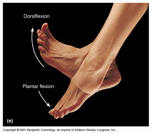
§ Abnormal toe motion: This would refer to a lack of toe push off (plantar flexion) or landing, or abnormal toe deviation (in or out).
§ Excessive pronation: Refers to excessive inward roll of the foot after contacting the ground. With excessive pronation the foot continues to roll in when it should be pushing off. This twists the foot, knee, leg and even the hip.
§ Excessive supination: Refers to inadequate inward roll of the foot after contacting the ground. This also causes strain on the foot, knee, leg and even the hip.
o Abnormal deviation of the knees. (Gait and single leg tests)
§ Restricted flexion or extension of the knees.
§ Medial or lateral deviation of normal knee motion during a single leg squat.
o Abnormal Leg alignment (femoral alignment)
§ Restrictions in internal or external leg motion (Femoral rotation).
o Abnormal hip or pelvic motion

Common Factors
On of the most common things I will see during gait analysis, is a lack of control as the individual brings their foot down (eccentric contraction): this motion is controlled by muscle called your dorsi flexors (tibialis anterior, extensor hallucis longus, extensor digitorum longus, peroneus longus). A person who has restrictions in these muscles is easy to hear when you are running. They will be the one slapping the ground as they run beside you. This is a significant finding, because it also means the person is not dissipating the shock of a normal stride. Instead they are directing the force into their muscles, which causes micro tears, inflammation and the formation of scar tissue. In fact, any deviation from a normal gait will have a similar effect somewhere in the kinetic chain, which is why all restrictions must be addressed for a complete resolution of this condition. These restrictions could be restrictions far from the feet themselves.
Treatment Strategies
Once the practitioner has determined (hypothesized) which areas are involved, they need to get into the restricted tissue to confirm where the adhesions are and release them. This will require that the practitioner use a considerable amount of tactile sensitivity. It is not just a matter of finding a tight spot and releasing it. The practitioner must literally feel what is going on in one layer of soft tissue over the adjacent layer.
This is achieved by having the patient perform specific movements/actions in conjunction with the treatment procedures. Each layer must be examined for motion, texture, tension, and function. By tissue motion I am referring to: how well the tissue glides over adjacent tissues, range of motion, elasticity, relative position, and even how it is affecting joint function and position.
When we take a closer look at the anatomy of your feet it becomes very obvious why testing each of these factors becomes so important. Let’s start with the bottom of your feet – the plantar surface. There are four layers of soft tissue structures on the bottom of your feet that should be addressed. Again, this is only one region of possible kinetic chain involvement.
Functional Anatomy
When you observe a deviation from normal motion patterns it is a direct indication of what anatomical structures are involved in a specific injury. This information tells the practitioner what primary muscles that perform the action could be involved (agonists), or what oppositional muscles (antagonists) could be a factor. This, combined with a whole body examination of kinetic chain relationships, provides the practitioner insight into what it will takes to resolve your particular case of Plantar Fasciitis.
First Layer of Muscles (Superficial layer)
The first layer of tissue is where we find the plantar fascia. It is very interesting that this layer is seldom tender or even that involved in Plantar Fasciitis, which usually involves the deeper structures.
o Flexor digitorum brevis: Flexes the toe joints (MP & PIP joints).
o Abductor hallucis: Moves the big toe (hallucis) away from the second toe (abduction), and assists in flexing the big toe.
o Abductor digiti minimi: Moves the little toe away (abduction) from the fourth toe, and assists in flexing the little toe.
Second Layer of Muscles
o Quadratus plantae (QP): This is an interesting muscle because it attaches to the tendon of the flexor digitorum longus (FDL). This muscle assists with flexion of the four lateral toes.
Note: At one end, the FDL originates under your calf muscles, and then its tendons go all the way down to insert under your foot on the four lateral toes. A tight FDL will affect the function of the QP.
Note: At one end, the FDL originates under your calf muscles, and then its tendons go all the way down to insert under your foot on the four lateral toes. A tight FDL will affect the function of the QP.
o Lumbricals: Four muscles with no bony attachment, they attach from the tendons of the FDL to the tendons of the EDL (extensor digitorum longus). These muscles help to flex and extend the toes.
Note: The function of these muscles will be affected by tension in both the FDL and EDL.
Note: The function of these muscles will be affected by tension in both the FDL and EDL.
Third Layer of Muscles
o Flexor hallucis brevis: Flexes the big toe (MP joint).
o Adductor hallucis: This two headed muscle moves the big toe inward (adduction).
o Flexor digiti minimi brevis: Flexes the little toe (MP joint).
Fourth Layer of Muscles
o Dorsal interossei: Four muscles that result in outward motion (abduction) of the third and four toes.
o Plantar interossei: Three muscles that result in inward motion (adduction) of the third, fourth and fifth toes (MP joints).
Plantar Fasciitis (PF) is a condition that does require a certain amount of in-depth analysis on the part of the practitioner. I have just shown you one aspect of the kinetic chain. In reality, each case of Plantar Fasciitis may present with the same symptoms but involve totally different structures. Therefore, the practitioner must individually examine the kinetic chain from your foot right up to your core. Then they must provide specific treatments and exercises based on individual needs. Here is an overview of how we approach each case of Plantar Fasciitis.
Implement Treatment Procedures:
o Next we Confirm our hypothesis on which structures are involved. This will be a continual process of concurrent diagnosis and implementation of procedures throughout the duration of the treatment appointments. The area of treatment focus may change depending on where the primary restrictions are found.
o There are several treatment procedures that can be effective in breaking up the adhesions formed along the Kinetic Chain. Some of my recommendations are: Active Release Techniques, Graston Technique, Registered Massage Therapy, and Fascial Manipulation.
Concurrent Exercise and lifestyle recommendations:
Exercise and lifestyle recommendations can be implemented before, during, and after treatment. Besides restoring the quality of the tissue by breaking up any restrictions it is important to become stronger and more flexible, and to build more power. This will help insure that the Plantar Fasciitis does not return.
Note: Most importantly once the adhesions have been removed it is exercise that will restore normal motion patterns and complete the process of tissue repair. With out the right exercises even if the adhesions are removed the probability of a reoccurrence in very high.
 If you would like more information or to purchase our books please go to www.releaseyourbody.com .
If you would like information about our clinic in Calgary Alberta please go to www.kinetichealth.ca.
(COPYRIGHT KINETIC HEALTH 2012 – ALL RIGHTS RESERVED)
If you would like information about our clinic in Calgary Alberta please go to www.kinetichealth.ca.
(COPYRIGHT KINETIC HEALTH 2012 – ALL RIGHTS RESERVED)
We offers prime services and solutions, with the goal of providing the best and latest technology in Plantar Fasciitis at highly competitive prices.
ReplyDeleteHi,
ReplyDeleteI am a student at Ashford University and I am currently working on my Senior Thesis research study project. I am looking at Plantar Fasciitis and the comparison among athletes vs gender. I was wondering if I am able to use your copyrighted photo at the top of this blog to put into my paper. If so, I will need some type of permission in text from you. My email is ggracejohnson@gmail.com and I hope to hear back from you soon.
Thanks,
Genevieve Johnson