Monday, October 25, 2010

Resolving Groin Injuries – Part 1

Groin injuries are a common athletic injury in both contact and non-contact sports. Injuries to the groin can occur from direct trauma in sports such ice hockey, basketball, football, rugby, and in non-contact sports such as gymnastics. In hockey, 10% of the injuries that occur each year are groin injuries. These injuries should be taken seriously because, once an athlete injures their groin, they are twice as likely as other players to incur the same injury again.
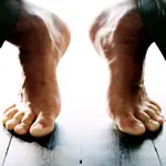
Anatomically your groin is the area where your upper thigh meets your pelvis (lower abdomen), essentially the crease or fold between these two areas. In reality, groin injuries involve a much larger area than just this. A groin injury can encompass an area that extends from your lower abdomen, to your pelvis, to your hip and inner thigh, and right down to your knee.

Since a groin injury can cover such a large anatomical region it can involve muscles, tendons, ligaments, joints, connective tissue (fascia), neurological, circulatory structures and even internal organs in the lower abdomen. In this blog we will limit our focus to musculoskeletal causes, except for a quick review on some conditions that must be ruled out (See differential diagnosis).

Differential Diagnosis
A differential diagnosis refers to the process of determining the probability of one condition over another. It is the process of making sure we are actually dealing with a musculoskeletal condition and not something more serious (pathological).
Fortunately most groin injuries are musculoskeletal injuries (mechanical problems) that will respond well to manual therapy and exercise. However, it is still important to evaluate other causes of groin pain (pathological causes) rather than assuming the problem is just a simple muscle strain. Some of the other problems that must be considered are:
· A hernia is a condition that must be ruled out whenever a groin injury presents itself. Minor hernias do respond to rest, ice, and range of motion exercises. More severe hernias require surgery (mesh reinforcement).
· The hip contains approximately eighteen superficial and deep bursae. Bursae are small pouches with slippery surfaces that reduce friction between two moving surfaces (increase gliding). The bursae are usually located in areas where muscles and tendons glide over bones. When the bursa becomes inflamed it loses the ability to decrease the friction between the moving surfaces. As inflammation increases, instead of providing ease of movement, they become a source of friction and pain. Chronic bursitis can be due to an underlying inflammatory condition. Examples of common sites of hip pain due to a bursa are the:
· Directly over the greater trochanter
§ Between gluteus maximus tendon and the greater trochanter (posterolateral prominence).
Fractures (Frank or Stress fractures)
· If the initial groin injury has been caused by trauma (in a contact sport) then possible fractures must be considered. The two most common sites for stress fractures are in the upper leg (femoral neck) and in the pelvis (pubic ramis). A simple X-ray is often all that is needed.
· An avulsion often refers to a bone injury where the tendon (which attaches muscle to bone) is torn away from its insertion point. Some of the possible sites of avulsions in a groin injury are:
§ ASIS (anterior superior iliac spine) – The site of the sartorius muscle attachment.
§ AIIS (anterior inferior iliac spine) – This is the site where the middle quadriceps (rectus femoris) attaches.
§ Ischial tuberosity – This is the site of attachment for your hamstrings.
· This condition is also known as gracilis syndrome. It is a repetitive stress injury that affects the pubic symphysis. This is an injury that can occur in runners, or sports that involve jumping or kicking. Osteitis Pubis can be increased after childbirth due to ligamentous laxity.
Nerve Compression
· Nerve compression can be a common problem associated with a groin injury. An example would be compression of a nerve called the femoral cutaneous nerve. When this nerve is compressed it creates a condition called Meralgia Paresthetica. This condition creates alter sensations on the outside of the hip.

In part two of Resolving Groin Injuries we will cover diagnosing a musculoskeletal groin Injury, including specific structures involved.
If you would like to more information or to purchase our books please go to www.releaseyourbody.com . 

If you would like information about our clinic in Calgary Alberta please go to www.kinetichealth.ca.

(COPYRIGHT KINETIC HEALTH 2012 – ALL RIGHTS RESERVED)

Wednesday, October 13, 2010

Ankle Stability – The Retinaculum Part 3


Injury To The Retinaculum

When functioning correctly, tendons glide under these retinacula without hindrance. With injury (trauma, repetitive strain), the retinaculum can become a site of tendon restriction, nerve impingement, and circulatory compression. Injury to the retinaculum will cause mechanical and neurological damage.
From a mechanical perspective, when tension is created between the retinaculum, and the structures that pass under them, a considerable amount of tension occurs. This tension can be a major problem since tension creates friction, which can cause micro-tears in the tissue, inflammation, and eventually adhesion formation. These adhesions inhibit relative motion, alter lower extremity biomechanics, and lead to a host of compensations.

From a neurological perspective, injury causes an alteration in neurological receptors (mechanoreceptors and proprioceptors). This leads to both ankle and foot instability. Instability of the ankle and foot creates abnormal motion patterns, compensations which can lead into numerous injuries throughout the body.

Treatment
Restrictions of the retinacula can be treated quite effectively with Manual Therapy (Active Release Techniques, Graston Technique, and Massage Therapy) and a series of corrective exercises. Treating with manual therapy involves breaking restrictions between the retinaculum and the tendon. Essentially the practitioner is restoring relative motion between the retinaculum and the tendons (and of course the muscles that the tendons are attached to).

The practitioners should also be focusing their treatment on the fascial lines of stress. Lines of stress in fascia are often created during injury in multiple locations not just at the site of pain. If these areas of fascial stress can be released, then normal fascial tension can be restored.

Restoring overall fascial tension, besides releasing adhesions between retinaculum and the soft tissues that pass under them, can have significant effects in resolving an injury.

Fascial interconnections are not theoretical entities; they are actual physical structures that have been mapped out. Researchers such as Thomas Meyers (Anatomy Trains) and Luigi, Carla, and Antonio Stecco (Fascial Manipulation) have spent decades researching these interconnections. During the last International Conference About Fascia at the University of Amsterdam, I had the privilege of listening to medical experts from around the world confirm this and related fascial research.

In my own clinical experience we have seen excellent results in improving ankle stability by removing adhesions at the retinaculum itself, but even better results when we work on restoring overall fascial tension

Next year I will be traveling to Italy (June and September) to do advanced training with Carla, and Antonio Stecco. I am sure that by next year I will have discovers even more about this fascinating subject.

Exercise
Exercise plays a significant role in the rehabilitation of a retinaculum injury. Strengthening and flexibility exercises are needed, but because a significant component of a retinatculum injury involves neurological receptors, balance and proprioception exercises are also essential for full recovery.
The following links are examples of exercises that we often recommend for out patients with injuries to the retinaculum (from Core Performance).

If you would like more information or to purchase our books please go to www.releaseyourbody.com . 

If you would like information about our clinic in Calgary Alberta please go to www.kinetichealth.ca.

(COPYRIGHT KINETIC HEALTH 2012 – ALL RIGHTS RESERVED)

Tuesday, October 12, 2010

Ankle Stability – The Retinaculum Part 2


In part two of this blog we continue by first covering the lateral and medial retinaculum and what structures pass underneath them.


Outside (Lateral) Ankle Retinaculum
Pain/Symptom pattern: Tension or a restriction in this area will often cause lateral ankle pain, altering both foot and ankle motion. This can easily lead to ongoing injury and a decrease in athletic performance.

Note: Peroneal retinacula are often injured during ankle sprains (inversion injury). Anytime there is persistent pain after an ankle sprain, a retinaculum injury should be considered. For more information on ankle sprains read my six part blog on Ankle Sprains (Inversion Sprain).

Inside (Medial) Ankle Retinaculum
Pain/Symptom pattern:
  • Restrictions of the flexor retinaculum are associated with what is known as Tarsal Tunnel Syndrome. In this syndrome people experience sensation of: Pain, tingling and altered sensation anywhere from the ankle, heel, bottom of the feet, to the toes.
  • A person with this syndrome will often experience an electric shock sensation, which travels into the foot, when they tap directly over the retinaculum. This is also known as Tinel’s sign.
Tarsal Tunnel Syndrome (TTS): Tarsal Tunnel Syndrome refers to compression of the posterior tibial nerve in the flexor retinaculum.
  • Persons with flat feet (fallen arches) are susceptible to TTS. Also any type of enlargement in the Tarsal Tunnel can cause this syndrome which includes swollen tendon, cysts, arthritic bone spur, varicose veins, or even inflammation in the surrounding area.
  • If this syndrome is left to progress it can lead to permanent nerve damage.
  • Conventional therapy can often be very effective in treating this syndrome (Rest, Ice, and Exercise). Active Release Techniques has developed some specific protocols the release the posterior tibial nerve in the Tarsal Tunnel.
In part three of Ankle Stability – The Retinaculum, we will injury and treatment.

If you would like more information or to purchase our books please go to www.releaseyourbody.com . 

If you would like information about our clinic in Calgary Alberta please go to www.kinetichealth.ca.

(COPYRIGHT KINETIC HEALTH 2012 – ALL RIGHTS RESERVED)

Monday, October 11, 2010

Ankle Stability – The Retinaculum Part 1


Usually when we think about ankle problems, we think about sprained ankles or a strained muscle, not something called a retinaculum. Yet these fascial structures play a significant role in a wide variety of chronic ankle problems.
So what is a retinaculum? From one perspective a retinaculum is a band of thick deep fascia that holds the long tendons of your ankle (those that cross the ankle) in place. Retinaculum also acts as a pulley system increasing mechanical advantage.

From the second perspective retinaculum are a major source of neurological receptors involved in balance and proprioception. Essentially retinacula have been hypothesized as key structures in spatial control for foot and ankle movements.

The following section is an overview of specific retinacula and what structures pass underneath them. As you look over the individual sections of the retinaculum also think of these areas as part of one large fascial interconnecting unit.

Retinacula do not exist as they are illustrated
Last year at the second international fascia conference in Amsterdam it became very clear to me that retinacula do not exist as they are illustrated in textbooks. There is a lot of interconnecting fascia that has to be removed before retinaculum look the way they are presented in text books. Research is now showing that these fascial connections (which are removed by dissection) are very important for both force transmission and neurological function.

Retinaculum Anatomy:
Front (Anterior) Ankle Retinaculum
Extensor retinaculum (2 parts)
o This structure holds in place tendons from the following muscles; tibialis anterior, extensor digitorum longus, extensor hallucis longus, and peroneus tertius.
o The deep peroneal nerve also passes under the retinaculum.
o The inferor retinaculum is shaped like a Y (once the entire surrounding fascia is removed) and has a lower and upper portion. The Y shape has the function of preventing “Bowstringing” of the tendons during ankle motion.
Pain/Symptom pattern: If there is a restriction in an extensor retinaculum, (front of the ankle) you may experience the following symptoms:
  • Localized pain or restriction on the front of ankle when running or walking. It is a very common symptom that I see with runners.
  • Tension can also alter the muscle firing patterns in the lower extremity. This can create a host of injuries and result in a substantial decrease in athletic performance
In part two of Ankle Stability – The Retinaculum, we will cover the lateral and medial retinaculum.

If you would like more information or to purchase our books please go to www.releaseyourbody.com . 

If you would like information about our clinic in Calgary Alberta please go to www.kinetichealth.ca.

(COPYRIGHT KINETIC HEALTH 2012 – ALL RIGHTS RESERVED)

Tuesday, October 5, 2010

Iliotibial Band Syndrome (ITBS) - Part 4


Treatment of Iliotibial Band Syndrome

The first thing practitioners must do in treating Iliotibial Band Syndrome (ITBS) is to perform a biomechanical analysis. In this process they evaluate the patients gait for alteration in movement patterns. This will give them an initial hypothesis as to which structures are involved. Refer to Parts 2 and 3 of this Blog to see what muscles are involved in what actions.

Then the practitioner will need to examine these areas (hands-on palpation) to confirm that there is a restriction present. The practitioner will feel an alteration in normal tissue consistency, it will feel ropy, rigid, and there will be a lack of tissue glide between adjacent structures. Once the area to be treated has been identified, manual therapy can begin. There are several forms of therapy that can achieve good results in addressing myofascial adhesions (Active Release, Graston Technique, Massage Therapy, and Fascial Manipulation).

Fascia and ITB Syndrome
When we evaluate a case of ITBS, we must also take into consideration all the fascial connections to the structures that are involved in performing and coordinating ankle, knee, and hip motion.

In the case of ITBS, some of the fascial connections we should consider are in the Lateral Line (see Anatomy Trains by Thomas Meyers).
The following is a short synopsis of the lateral line. I have also included a link to the Anatomy Trains dissection video about the Lateral Line – Thomas Meyers Lateral line Video.
Lateral Line
· Beginning at the foot with the peroneal muscles, fascial interconnections travel up the outer leg to just below the knee (fibular head). This fascia then connects directly into the lower IT band into the deltoid complex (gluteus maximus, gluteus medius, and tensor fascia latae).
· The fascia then connects to the pelvis (iliac crest), which connects into the abdominal muscles (internal and external obliques), and then into the quadratus lumborum which connects into the ribs and the spine.
· The fascia then travels up between the ribs (intercostals muscles) and continues up the body to connect into the fascia of the neck (SCM, splenicus cervicis, and scalenes).

Once you see these videos, you will find it very hard to dispute the importance of these fascial connections.

Bottom Line: You can try treating ITBS with all the right techniques (ART, Graston, Massage etc.) and find that you still have not resolved a chronic ITBS problem if you do not consider, and treat, the fascial interconnections.

The key is to any therapy is to address all the structures involved in the kinetic chain.
Treatments need to be specific and based upon the individual requirements of each person. Treatment should not be a cookbook approach. Practitioners need to find the specific tissues that are restricted and physically work them back to their normal consistency.

Exercise is Essential
Often getting professional help in the way of manual therapy is essential to get a complete resolution of ITBS, but just as important is exercise. Without the right exercise program the probability of this condition returning is very high.
There are three important areas which must be addressed when prescribing exercise routines for ITBS, strength, flexibility, and balance.

Why strengthening exercises are essential:
Every time you injure yourself, your body lays down new tissue to repair itself. The new tissue is initially very fragile, thin, and easily torn or re-injured. Strength or weight training places stress upon these new tissues, causing them to go through a process of remodeling. In this process, the new tissue literally converts from one type of collagen to a different type which is up to 10 times thicker and 10 times stronger. However, this collagen conversion only occurs when you apply continued stress upon the tissue as you do in weight- and strength–training exercises.

Exercise example: Because the iliotibial band is formed from the deltoid complex, having strong balanced hips is essential for a full resolution of ITBS. One of my favorite exercises to increase hip strength is the Bulgarian Split Squat.

Why Flexibility exercises are essential:
During the regenerative or repair phase of an injury, your body creates and lays down collagen to repair the injured area. When the injured person performs the correct stretching exercises, the majority of new tissue will be laid down in the same direction as the tissue that is being repaired - thereby allowing this tissue to properly perform its function.
Exercise example: We suggest that patients combine stretching with self myofascial release (foam rollers). The following is an example of an ITB stretch, and a foam roller video you will find very useful.

Why balance exercises are essential:
Proprioceptive or balance training is a fundamental requirement that should not be ignored in Rehabilitation Therapy. Your ability to balance depends on feedback from your auditory, visual, proprioceptive (sense of body position), and vestibular systems (relating to the sense of equilibrium). All of these systems must be trained to achieve optimal results. As with all injuries your nervous system is often affected. Training these systems, ensures a greater chance of a complete recovery.

Exercise example: The follow exercise is an example of the type of exercises that we prescribe to our patients. This is the Squat - Single leg balance exercise from Core Performance.


If you would like more information or to purchase our books please go to www.releaseyourbody.com . 

If you would like information about our clinic in Calgary Alberta please go to www.kinetichealth.ca.

(COPYRIGHT KINETIC HEALTH 2012 – ALL RIGHTS RESERVED)