Showing posts with label Active Release Techniques. Show all posts

Wednesday, November 20, 2013

Treating Bunions

Bunions (Hallus Abducto Valgus) are a common foot problem that affects the joint at the base of the big toe (first metatarsophalangeal joint). In Latin “bunion” means enlargement, while “hallux abducto valgus (HAV)” refers to a bending inwards of the big toe as seen on the image. As you see, the big toe bends in towards the other toes while the bone behind it (1st metatarsal) pushes outward. This creates a considerable amount of stress on the joint (first metatarsophalangeal joint). Due to this bending inwards, a sharp angle at the big toe joint is created, resulting in the formation of a bunion. Initially, this enlargement is composed of swollen tissue which becomes irritated by any external pressure (for example tight shoes). Eventually this swollen tissue thickens to form a very large lump or bunion.

There is an obvious relationship between bunions and shoes, since bunions do not occur in cultures that go barefoot. High heels, pointed shoes, ballet shoes, excessively tight shoes, and even cowboy boots often lead to the development of bunions. 


Anatomy and Biomechanics

From a biomechanical perspective, bunion formation creates a cycle of dysfunction. As the bone behind the big toe (1st metatarsal bone) moves outwards, the inner arch of the foot becomes unstable and starts to collapse. This instability, or lack of support in the arch, increases stress on the angle at the point where the bunion is forming. This stress accelerates the formation of the bunion, which in turn further destabilizes the arch of the foot. To truly deal with this problem, you must address both the foot instability and joint angle.

In addition to the stresses caused by poor shoes, simply walking with your feet in a "turned out" position can also lead to bunion formation from the stress it puts on two particular muscles, the adductor hallicus and the abductor hallicus.

The adductor hallicus is an interesting muscle which is shaped like the number seven. The adductor hallicus transverses from several of the lateral toes into your big toe. When the adductor hallicus contracts, it pulls the big toe towards the 2nd toe. When the adductor hallicus muscle becomes tight and restricted, it continually pulls the big toe towards the second toe (even without contracting). The adductor hallicus tends to become restricted in individuals who excessively pronate or walk with their feet turned outwards (a huge percentage of runners and dancers).

This pulling action of the adductor hallicus disrupts a key balance in muscle tension, which normally keeps the big  toe in a neutral or straight position. This balance occurs between the adductor hallicus and the abductor hallicus muscles. The abductor hallucis normally resists the pulling action of the adductor hallicus. The abductor hallucis runs from your heel (calcaneus) to your big toe (proximal phalanx).

Constant pulling from the adductor hallicus weakens and overstretches the abductor hallucis. Without the appropriate counter-balancing action of the abductor hallucis, the big toe moves inward, and bunion formation accelerates.

Treatment

The objective of non-surgical treatment is to prevent the progression of the bunion by correcting the biomechanical stress on the foot, by realigning the joint as much as possible, and by increasing the intrinsic strength of the foot. We achieve this by using soft-tissue tissue techniques such as Active Release Techniques to break adhesions that form in the adductor hallicus muscle or other affected structures.

We use taping help bring the big toe back into a neutral position.  We have our patients perform a series of strengthening, stretching, and self-myofascial release exercises.  The selection of exercises will vary greatly between different patients, and is dependant on which areas of their kinetic chain has been affected.

Taping a Bunion

Bunion taping can help reduce pain and discomfort of a bunion by helping to realign the joint taking pressure off the bunion.


Exercises
The following two exercises are examples of exercises that we recommend for Bunions. Please note, this is just a sample of our exercises, the actual exercise routine will vary depending on each individual case.


Increasing Dorsi Flexion A decrease in dorsi-flexion causes excessive pronation of the foot, the knee to move in (knee valgus), and internal rotation of the the leg (femur).


Increasing Big Toe Mobility Having mobility in the joints of the big toe is essential for normal gait.


Do you want more information about this, and other topics. Our books (eBooks and hard-copy) provide more information about soft-tissue injuries, rehabilitative exercises for injury recovery, and how to use these to activate and restore all the structures of your kinetic chain.  

To purchase our internationally best-selling books, visit www.releaseyourbody.com  


For information about our clinic in Calgary, Alberta, please go to www.kinetichealth.ca. Or call us at 403-241-3772. 

(COPYRIGHT KINETIC HEALTH 2013 – ALL RIGHTS RESERVED)

Friday, February 24, 2012

Patellar Tendonitis/Tendinosis or Jumper’s Knee

The patellar tendon links the kneecap (patella) to your shin bone (tibia). The quadriceps muscle and the quadriceps tendon allow your knee to extend.

Tendonitis refers to inflammation of a tendon. Tendonitis in the knee is commonly caused by activities that shorten the quadriceps, and that transfer force directly to the tendons of the knee. This force causes friction and inflammation of the tendons, making it difficult and painful to run, walk, or perform any weight-bearing motion.

Tendonitis of the knee is common in ball players, runners, cyclists and triathletes. It is also common in the elderly, or in extremely inactive individuals. Untreated tendonitis can eventually lead to tearing and rupture of the tendon.

Traditionally, tendonitis/tendinosis is treated by icing during the acute stages of the injury, reducing physical activities, and by the consumption of non-steroidal anti-inflammatory drugs (NSAIDs). These are short-term treatments that should only be applied during the acute stages of the injury. Most of these treatments are limited in their effects, and they provide only symptomatic relief – they act to reduce inflammation – but do not address the underlying biomechanical problems that caused tendonitis/tendonosis.

In addition, the long-term consumption of non-steroidal anti- inflammatory medications has several detrimental side-effects including gastrointestinal problems, ulcerations, and internal bleeding.

Treating Patellar Tendonitis with Soft Tissue  Therapy

Patellar Tendonitis, or Jumper’s Knee, usually responds extremely well to Soft Tissue Theapy (ART, Graston, Register Massage Therapy, Fascial Manipulation). The pain caused by Patellar Tendonitis is usually felt between your kneecap (patella) and its attachment point on your shin bone (tibial tuberosity of your tibia). Essentially, the entire knee capsule must be evaluated and any restrictions that are found need to be removed.

It is equally important to remove any restrictions in the quadriceps muscle since muscle fibers from the quadriceps combine at the knee to form the patellar tendon.

In addition to removing adhesions and restrictions, you should also address the following key factors to obtain a full resolution of this condition:
  • Strengthen Your Knee’s Kinetic Chain – It is important to perform exercise routines that strengthen and support all the elements of the knee’s kinetic chain even when that exercise does not appear to directly affect knee function.
  • Address Muscle Imbalances –The muscles that form the quadriceps femoris group can be incredibly strong. This strength has to be counter-balanced by an equivalently strong hamstring. When there is a large imbalance in strength between these two muscle groups, increased force is placed on the patellar tendon which attaches to the knee. Muscle imbalances in the hips and core also affect these structures, and need to be addressed for long-term resolution of the knee injury.
  • Reduce Obesity – Research has shown that carrying an extra 10 to 20 lbs of weight will greatly increase the stress on the patellar tendon. Weight management can be an important factor in resolving this condition.
  • Manage Your Training Intensity – It is important to pay attention to the onset of your patellar tendonitis. If you find that the condition occurs suddenly after an increase in training intensity, then you should be careful and restrict your increases to 5%-10% per week. Any increases above this level increases the probability of another injury.
  • Get Enough Rest – It always amazes me that people are so unwilling to give their body sufficient time to heal. Rest is a critical component of the healing cycle, so give your body enough time to heal between your workouts.
  • Wear Proper Footware – Good footwear is essential to the healing process, especially if you already have gait imbalances (over-pronation or supination).
By combining Soft Tissue treatments (ARTGraston, Register Massage Therapy, Fascial Manipulation)  with exercise, and by following the above recommendations, you can usually expect substantial functional improvements in your ability to perform your daily living tasks (climbing stairs, sleeping, running, and jumping) as well as a reduction in your pain.


If you would like more information or to purchase our books please go to www.releaseyourbody.com . 

If you would like information about our clinic in Calgary Alberta please go to www.kinetichealth.ca.


(COPYRIGHT KINETIC HEALTH 2012 – ALL RIGHTS RESERVED)

Tuesday, February 14, 2012

How Abnormal Motion Patterns Cause Shoulder Injuries


Many shoulder problems are caused by abnormal motion patterns that occur as the body attempts to continue performing daily living activities while compensating for the effects of previous injuries. Some of the most common abnormal motion patterns that we see are caused by:
  • Shoulder Joint Instability
  • Abnormal Movements of the Shoulder Blade
  • Restrictions in Internal Shoulder Rotation
Shoulder Joint Instability
Research has shown that extended periods of shoulder instability (glenohumeral instability) can start a cycle of micro-trauma and secondary impingement syndromes which often result in chronic shoulder pain. Practitioners will often find this type of instability during shoulder examinations and translation tests.
Anterior shoulder instability (laxity of the anterior capsule) is often related to problems in the posterior shoulder capsule. This combination of problems can have a considerable impact upon the structures of the shoulder’s kinetic chain
When the posterior shoulder capsule becomes tight, it affects the IGHL (inferior glenohumeral ligament). This ligament helps to maintain your shoulder’s position in the joint and acts as a supporting hammock or sling for the humeral head. When your IGHL does not function the way it should, your arm (humeral head) changes position, which can then result in a variety of impingement syndromes.
Abnormal Movements of the Shoulder Blade
Numerous studies have shown that abnormal motion patterns of the shoulder blade (Scapular Dyskinesis) can result in a variety of shoulder impingement syndromes. These alterations in muscle activity are often overlooked by clinicians when reviewing patients who have been diagnosed with a rotator cuff injury.
Abnormal shoulder blade motion typically causes an alteration in the muscle firing patterns of the:
These abnormal motion patterns affect the patient’s ability to bring their shoulder forward or backward (protraction and retraction). A lack of backward motion (retraction) often leads to hyperangulation - a risk factor that can lead to shoulder impingements.
I believe that it is extremely important to restore normal motion patterns for the shoulder blade, since the presence of abnormal motion patterns substantially increases the probability of an injury reoccurring.
A practitioner who tests for and discovers alterations in normal shoulder motion can apply the appropriate Soft Tissue procedures and exercises to solve this problem.
Restrictions in Internal Shoulder Rotation
An increased risk of shoulder impingements is often associated with restricted internal rotation of the shoulder. This is especially true when an action requires the shoulder to be both flexed and internally rotated.
In this position, increased pressure is experienced between the insertion of the supraspinatus muscle and the acromion or coracoacromial ligament. This area often becomes the focal point of an impingement. For example:
  • The supraspinatus muscle is a rotator cuff muscle that raises the arm to your side.
  • The acromion is a part of the shoulder blade (scapula) that lies above the shoulder joint. The acromion articulates with the clavicle to form the acromioclavicular joint which is a common restriction point.
  • The coracoacromial ligament is a strong triangular band on the shoulder blade, which with the coracoid process and the acromion, forms a vault for the protection of the head of the humerus.
Consequently, in cases where restrictions in internal rotation are noted (common with rotator cuff injuries), the practitioner should always look for and address impingements at these important focal points.

If you would like more information or to purchase our books please go to www.releaseyourbody.com . 

If you would like information about our clinic in Calgary Alberta please go to www.kinetichealth.ca.


(COPYRIGHT KINETIC HEALTH 2012 – ALL RIGHTS RESERVED)

Thursday, February 9, 2012

Effects of Excessive Pronation on the Knee

For an example of the interactions between the structures of the knee’s kinetic chain, let us take a look at a person whose foot is excessively pronated (rolled inwards). This pronation causes the person’s foot to flatten out during normal walking. This flattening then causes the tibia to rotate inwards (medially) and the femur to rotate outward (laterally).


These actions place a considerable amount of stress on the knee, eventually leading to friction, inflammation, altered motion patterns, and injury of the soft- tissues of the knee. Thus, a problem that started at the foot ends up causing abnormal hip and femur rotation, which in turn leads to knee problems.

From a therapy perspective, it is possible to achieve moderate success by treating just the immediate structures of the knee. However, in order to truly resolve the problem, we should also treat those structures that were the original cause of the excessive pronation – that is, the structures in the knee’s kinetic chain. For example, restriction in any of the following structures may be the actual cause of the excessive pronation:
  • Peroneus longus and peroneus brevis muscles help you to point your feet and aid in eversion (rolling inward) of the foot when walking or running.
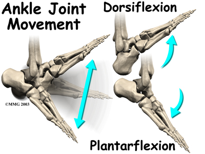
  • Tibialis anterior lets you bend your foot upwards (dorsiflexion) and also helps to invert the foot (roll outwards) when you walk. Proper inversion of the foot is an important part of a normal gait pattern.
  • Abductor hallucis, this muscle is responsible for flexing the big toe and allows your big toe to move laterally (sideways). This is important since a normal walking/running stride requires us to push-off with our big toe.
  • Flexor hallucis brevis, this muscle is responsible for flexing the big toe and for supporting the medial arch of the foot.
  • Flexor hallucis longus, this muscle is responsible for flexing the big toe, supinating the ankle (turning inwards), and in pointing your foot (plantar flexion).
Restrictions in any of these structures can cause excessive pronation, which in turn leads to hip restrictions, and subsequent knee problems.

Obviously, in such situations, treating just the structures in the knee will not resolve the knee problem. Instead, the practitioner must treat the knee, and then, based on the biomechanical analysis, treat all other affected structures in the knee’s kinetic chain. The knee problem will only be resolved when restrictions in all these affected structures are removed.

Exercise
For every restriction that occurs, an altered muscle-firing pattern is also created. These dysfunctional movement patterns will still remain after the restriction (adhesion/ scar-tissue) has been removed. Only a corrective program of exercises will re-establish a normal motion pattern by retraining these structures to properly work together. This is why it is essential to combine the removal of the adhesions with appropriate and specifically designed exercise protocols.

Bottom line, as good as any therapy is without rehabilitative exercises the problem will most likely return.


If you would like more information or to purchase our books please go to www.releaseyourbody.com . 

If you would like information about our clinic in Calgary Alberta please go to www.kinetichealth.ca.


(COPYRIGHT KINETIC HEALTH 2012 – ALL RIGHTS RESERVED)

Wednesday, February 1, 2012

Medial Ankle Pain - Dancer’s Tendonitis


An injury to the Flexor Hallucis Longus (FHL) tendon causes medial ankle pain or pain on the bottom of the foot. FHL injury is a condition that is often overlooked or misdiagnosed. This injury affects dancers, runners, soccer players, and any other athlete who performs repeated, propulsive forces, or jumping. Injury of theFlexor Halicus Longus
muscle is sometimes called “Dancer’s tendonitis” but it is not limited to just dancers. Image
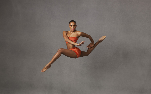
Alvin Ailey American Dance Theater dancer Constance Stamatiou.


The Flexor Hallucis Longus (FHL) muscle allows you to point your big toe (plantar-flexing your big toe) and stabilizes the Subtaler Joint. The Subtaler Joint is located between two bones in your ankle - the Talusand the Calcaneus. The Subtaler Joint allows movement of the heel toward the medial plane (inversion) as well as movement of the heel towards the lateral plane (eversion).

Anatomy/Biomechanics of Medial Ankle Pain
The Flexor Hallucis Longus muscle is located deep under your calf muscles (the most lateral muscle of the deep compartment). The FHL originates on the lateral lower leg (distal 2/3 of Fibula). It then travels at an oblique angle (crosses the posterior surface of Tibia) down towards the medial ankle (posterior surface of Talus) and travels under a section of the heel bone (Sustentaculum Tali of Calcaneus). The FHL then passes under the sole of the foot (between the two heads of the FHB) and inserts into the base of the big toe (base of the Distal Phalanx of Hallax).

As mentioned the FHL Tendon curves around a structure called the Sustentaculum Tali. TheSustentaculum Tali is a bony section on the inside of the heel bone (Calcaneus). This is often a site of friction and irritation for the FHL tendon. The FHL tendon also travels between the twoSesamoid bones just behind the big toe (Metatarso-Phalangeal Joint). The sheath that surrounds the FHL tendon is often irritated.

Causes and Presentation of Medial Ankle Pain
The repetitive motion of pushing-off with your foot (plantar flexion) during dance, running, soccer, and jumping can cause injury to the FHL.

Injury to the FHL tendon and muscle can present in a variety of ways - sometimes involving inflammation and sometimes not. Injury to the tendon without inflammation is refer to as a “Tendinopathy”. Tendinopathy refers to a degenerative lesion in the tendon without affecting the tendon sheath that surrounds the tendon.

When FHL inflammation (tendonitis) is present in the foot, it usually occurs in one of the following three locations.
2. At the “Knot of Henry” – a section just behind the big toe (first metatarsal) where the FDL muscle crosses the FHL tendon.
3. Just behind the big toe by the Sesamoids bone.
o A Sesamoid bone holds its tendon away from the center of the joint and acts to increase mechanical force.

When the FHL tendon becomes nodular, a condition called Hallux Saltans can develop. Hallux Saltans is similar to trigger finger in the hand, except it occurs in the big toe. Triggering of the toe occurs when the nodular thickening of the tendon snaps through the fibro-osseous tunnel. This causes a jerking motion, much like a trigger finger.

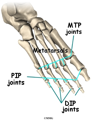
If not addressed Hallux Saltans can contribute to the progression of an additional condition called Hallux Rigidus. Hallux Rigidus means "stiff great toe". Hallux Rigidus is the second most common disorder of the first MTP joint. The most common injury is a bunion - otherwise known as Hallux Valgus.


Special Considerations For The Dancer
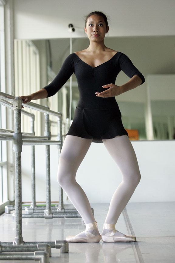
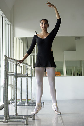
Injury to the FHL in dancers is often caused by the repetitive motion of changing position from a plié position to a relevé position. This action produces a force that is 10 times the dancer’s body weight.

Ideally, for greater stability and increased propulsion, the foot should be in a supinated position at the heel during push-off. For a dancer, any action that causes a reduction in plantar flexionmotion can create an FHL injury. Biomechanically, a lack of plantar flexion leads to a prolonged pronation position of the foot when pushing off in the Propulsion Phase.
In dancers, the FHL tendon is often compressed while performing a relevé position and is over-stretched while performing a plié postion. In such a case, the dancer will feel posterior medial ankle pain when performing the plié. (Photo - Dance Theatre of Harlem).

Diagnosis and Imaging - FHL Injury
X-rays for this condition are not very specific. X-Rays are good for ruling out fractures (Calcaneus, Distal Medial Malleolus, or Os Trigonum) that may cause an impingement of the FHL tendon. It is important to note that X-Rays will not provide a definitive diagnosis of FHL injuries.
A good history is much more definitive, especially when accompanied by a full physical examination and an MRI. MRI imaging is an excellent resource for showing damage to tissue fibers, inflammation, and swelling (edema).
If the MRI is not available or is too expensive, a CT scan (Computed Tomography) can prove to be a useful alternative.

Treatment
In most cases, FHL syndrome (involving partial tears, tendinosis, or inflammatory conditions) responds well to conservative care. However, a complete tear of the FHL tendon may require corrective surgery since serious cases of FHL injury could end an athlete’s career.

Conservative care includes:
· Ice or heat depending on stage and type of injury (inflammation or not). See our Icing andHeating blog.
· Reduction in activity. Usually, some modification in all activities is necessary.
· Supports (crutches or walker boot) can be beneficial.
· Taping – Kinesio Taping. At Kinetic Health we use SpiderTech Kineso-taping.
· Soft-tissue and joint mobilization (Active Release, Graston Techniques, Massage, and Fascial Manipulation).
· Inflammatory strategy implementation (medication for no longer than seven days). See ourblog about Reducing Inflammation.
· Exercises for increased strength, flexibility and balance. Go to www.releaseyourbody.comfor exercises you can use to treat this condition.
· Biomechanical corrections (footwear or Orthotics) .
· A gradual return to activities.

If you would like more information or to purchase our books please go to www.releaseyourbody.com . 

If you would like information about our clinic in Calgary Alberta please go to www.kinetichealth.ca.


(COPYRIGHT KINETIC HEALTH 2012 – ALL RIGHTS RESERVED)