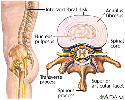
Spinal Disc Degeneration
Disc degeneration is part of the normal aging process.
As we age, our discs begin to shrink due to loss of fluid within the discs.
This loss of fluid in the disc leads to a decrease in the normal height of the
disc, thereby decreasing the disc’s ability to absorb shock.
The lack of shock absorption by the discs causes
increased stress on the facet joints (a gliding joint between each vertebra) of
the spine and results in facet joint degeneration.
These changes may eventually cause increased pressure
on the nerve roots (nerves that exit from the spinal cord) and may result in
sciatic-type pain down the arm or leg). This condition is often referred to as
Degenerative Disc Disease.
Disc Herniation, Protrusion, Prolapse, & Extrusion
A
disc protrusion (also known as a disc bulge) occurs when the inner material of
the disc starts to push out through the outer wall of the disc, creating a
bulge in the disc. In most cases this disc bulge is completely symptomless, and
causes no pain or lack of function. In fact, most individuals over the age of
forty have disc bulges.
Problems occur when these disc protrusions start to
fragment or tear. A herniated disc occurs when the inner material of the disc
(the nucleus pulposus) starts to push through the outer fibres of the disc (the
annulus fibrosus). Most disc herniations occur at the lower levels of the
spinal column.
When
the outer layers of a disc rupture, the inner centre of the disc may move out
and press upon a nerve. This condition is known as disc prolapse or protruding
disc. In such cases, the material inside the disc can sometimes extrude into
the vertebral (spinal) canal.
In
rare cases a severe prolapse will press on the nerves which control bowel and
bladder function, resulting in severe muscle atrophy. These are rare events and
are considered to be surgical emergencies. The majority of disc prolapses do
not fit into this category. In yet other cases, a disc may extrude right
through the outer fibres of the disc, and a piece may break off completely.
When this occurs, the extruded piece of disc can
interfere with the function of the nearby nerves. This condition – sequestered disc – requires surgical intervention if it is causing neurological
dysfunction; it is a problem that cannot be resolved with manual therapy.
However, the most important point to be made is that
most cases that involve a disc bulge or protrusion do not require surgery. In
fact there are a couple of common myths about disc protrusions that we should
consider:
- The first myth is that the presence of a large disc protrusion – often seen on MRI or CAT scan images – is an indication that this problem cannot be resolved with conservative care (non- surgical). In reality, research is showing the exact opposite to be true. The larger the disc protrusion, the greater the reduction in protrusion size after conservative treatment.
- The second myth is that the extruded and sequestered disc fragments are less likely to resolve than the contained protrusions. In actuality, the migrating fragments actually resolve more frequently and faster than the contained protrusions. The reason for this; the larger the disc protrusion, the greater the degree of inflammation around the protrusion. Once the disc fragments have broken off and inflammation has decreased around the fragments and disc decreases, the body is able to reabsorb the fragments easily.
Treating and Resolving Disc Injuries
Initially, disc injuries can feel incredibly painful,
and it may seem that surgery is the inevitable outcome. Fortunately, the
majority (95%) of patients suffering from disc injuries do NOT need surgery.
However, you will need to be patient as you work through the resolution
process.
The actual time required to resolve a disc injury
varies based on the severity of the injury, length of time the problem has
existed, your age, and your current and historical physical condition. To
effectively resolve a disc injury, it is necessary to:
- Remove any type of mechanical restriction that is causing stress upon the disc.
- Avoid all physical stresses that are perpetuating that problem. Anything that can be done to remove biomechanical stress from the back can benefit the patient. This includes:
Icing and Anti-inflammatories during the Acute Stage:
During the acute stage of the injury,
anti-inflammatories and ice are useful, but only provide relief at a
symptomatic level. During the initial stage of injury:
- Perform ice massage on the affected area for at least the first 72 hours to reduce pain, inflammation, swelling, and muscle spasms.
- Rest for a maximum of two days during the acute stage of the injury. In fact, you may find that resting is about as much as you can manage!
- Return to as many of your normal, daily living activities as you can manage after the first 48 to 72 hours. This will increase the speed of your recovery.
Performing the Activities of Daily Living:
With a
disc injury, you will have to find a way to perform your daily living
activities without aggravating your condition or increasing the resulting pain. This may mean that you must:
- Reduce your range of motion as you perform each task.
- Move more through your hips rather than through your back.
- Brace your core before lifting or moving any object
- Avoid repetitive actions that are performed in a flexed or bent- over spinal position since this can result in increased disc damage. This is especially true if you are lifting heavy loads from this position.
- Take frequent rest breaks. If you have a sedentary job (sitting at a desk), then take active breaks to walk around, and move all your limbs. If you have an active job, then slow down, sit, and relax for a few minutes.
- Listen to your body. If it says that a task is beyond your current capability, then stop before you cause further injuries which can change an acute problem into a chronic problem.
Using Soft Tissue Therapy and Spinal Manipulation
Any
injured muscle, joint capsule, ligament, tendon, fascia, or connective tissue
restriction can cause changes in motor patterns (neuromuscular imbalances)
which then directly affect spinal stability. The specific physical restrictions
that need to be addressed in a disc injury will vary depending on the
individual. However, the removal of any type of mechanical restriction is of
great benefit for any type of disc injury. The practitioner will need to
palpate the affected areas and conduct appropriate biomechanical analysis in
order to determine exactly which areas need to be addressed.
Imbalances
in muscular tension increase stress on the spinal discs, which then perpetuates
or causes further injury. Fortunately, those spinal imbalances can be addressed
with manual procedures.
Good results are obtained when these procedures are
combined with dynamic exercises that re-establish normal motor control.
For
example, soft tissue therapies (ART, Graston, Registered Massage Therapy, Fascial Manipulation) can be used to release restricted
structures around the discs, such as:
- Multifidus Muscles (Deep posterior extensors and stabilizers of the spine.) Research has shown that a disc injury can reduce neurological input to the multifidus muscle. This reduction in neurological input can lead to muscle wasting of the Type 1 and Type 2 fibres. ART procedures are able to help restore normal multifidus function.
- Abdominal Muscles and Extensors of the Back - An imbalance between the abdominal muscles and extensors of the back can affect muscle strength, lumbar lordosis (curvature of the lumbar spine), sacral angle, and spinal stability. Any imbalances between these antagonistic structures causes increased spinal tension and spinal instability, and places stress on the disc. ART protocols in combination with the proper exercises are very effective at resolving these imbalances.

If you would like information about our clinic in Calgary Alberta please go to www.kinetichealth.ca.
(COPYRIGHT KINETIC HEALTH 2012 – ALL RIGHTS RESERVED)