Showing posts with label Dr Brian Abelson. Show all posts

Thursday, June 28, 2012

Exercises for nerve compression syndromes of the upper extremity


In the last few weeks we have produced three videos that provide you with exercises for three different types of upper-body nerve compression syndromes.  Each video is directed towards a specific nerve entrapment syndrome: Median Nerve Entrapment, Radial Nerve Entrapment,  or Ulnar Nerve Entrapment. We recommend that you use these nerve-flossing and tensioning exercise videos in conjunction with the exercise routines in our book “Release Your Kinetic Chain from the Shoulder to Hand”. This combination of exercises and techniques will provide with the most effective and rapid means to recover with these nerve entrapment syndromes.


Nerve Distribution Chart
Us this chart to help identify which nerve compression syndrome is predominant in your hand(s).


This diagram is derived directly from our internationally best-selling book - Release Your Pain: 2nd Edition.  Use this diagram to help aid in identifying which nerve is affected in your upper body and arm.

Exercise Videos for Releasing Nerve Entrapments

Select and use the appropriate exercise video for your nerve entrapment syndrome.

Median Nerve

If you have a Median nerve entrapment (Carpal Tunnel or other nerve entrapment syndromes), then use the exercises in this video to floss, mobilize, and release the Median nerve from its surrounding tissues.


Radial Nerve


If you have a Radial nerve entrapment syndrome, then use the exercises in this video to floss, mobilize, and release the Radial nerve from its surrounding tissues.


Ulnar Nerve

If you have Ulnar nerve entrapment syndrome, then use the exercises in this video to floss, mobilize, and release the Ulnar nerve from its surrounding tissues.


(COPYRIGHT Dr. Brian Abelson 2012 – ALL RIGHTS RESERVED)

Exercises for the Shoulder to Hand


This book is especially useful for people recovering from injuries to the shoulder, arms, elbow, wrist, and hands.   Use the exercises in this book to rehabilitate, strengthen, and restore the function of muscles from your Shoulders to Hands. This is the second volume in the Release Your Kinetic Chain series of books. 


  • ndex=1&feature=plpp_videohttp://www.youtube.com/watch?v=oeV9IhFFpEk&list=UU4N_ZSgXi81y8dOVRDEFl6Q&index=15&feature=plpp_videohttp://www.releaseyourbody.com/release-your-pain-2nd-edition

Sunday, February 19, 2012

Achilles Tendonitis or Tendinopathy


About the Achilles Tendon
The Achilles Tendon is the strongest and largest tendon in the body. It is extremely vulnerable to injury due to its limited blood supply and the numerous forces to which it is subjected.

The Achilles Tendon is known as a co-joined tendon. This tendon joins directly into the calf muscles (gastrocnemius and soleus). The Achilles Tendon transmits the force generated by the calf muscles to produce the push-off required for walking, running, and jumping.

The area of the Achilles Tendon (approximately 2 to 6 cm above its insertion into the calcaneus) is very dense and under constant tension; consequently, this area has the poorest blood supply, which makes it extremely susceptible to injury and very slow to heal when it is injured.

The calf muscles associated with the Achilles Tendon are composed of several layers of muscles, with the large gastrocnemius and soleus muscles being the more superficial. Under these muscles is a deeper layer of three muscles – the tibialis posterior, flexor hallucis longus, and flexor digitorum longusInjuries, restrictions, or adhesions in any of these tissue structures can directly affect the function and strength of the Achilles Tendon.

Kinetic Chain Structures of the Achilles Tendon

Other structures of the Achilles Tendon’s kinetic chain that are commonly involved in Achilles Tendon injuries include:
  • The hamstrings, which are a group of muscles that include the biceps femoris, semitendinosus, and semimembranosus. Tension in these muscles causes increased stress upon the muscles of the lower leg.
  • The tibialis posterior, which lies deep within/to the calf muscles. This muscle inverts the foot (turns the foot inwards) and plantar flexes the foot (helps you to point your toes down).
  • The popliteus muscle, which lies deep behind the knee and is involved in medial knee rotation. When it is restricted, it may place increased stress upon the lower extremities.
  • The soleus muscle, which is a powerful plantar flexor of the foot and gives you the ability to rise up on your toes.
  • The flexor digitorum longus which works to flex toes 2 thru 5. It also helps to plantar flex the foot.
  • The flexor hallucis longus, flexor hallucis brevis, and the tibialis anterior muscles, which are all involved in cases of increased pronation and hyperpronation.
  • The plantaris muscle, which inserts into the middle one third of the posterior calcaneal surface (heel bone), just on the inside of the Achilles Tendon. This muscle assists in plantar flexion of the foot and is also involved in flexion of the leg.
What is Achilles Tendonitis or Tendinopathy
 
The term Achilles Tendonitis (“itis” implying the presence of inflammation) is commonly used to describe tenderness, pain, and swelling in the area just above the heel bone (2 to 6 cm above the calcaneus). A more accurate term would be Achilles Tendinopathy which identifies the presence of both tendonitis (inflammation) and tendinosis (small tears in surrounding tissue).

In our clinic, we typically see three types of injuries to the Achilles Tendon – paratenonitis, tendinosis, and rupture of the tendons:

Achilles Tendonitis/Paratenonitis: This injury is commonly known as Achilles Tendonitis and describes an inflammation of the paratenon - a sheath surrounding the Achilles Tendon. Paratenonitis is often caused by overuse or repetitive strain and commonly occurs in triathletes and runners.

Tendinosis: Refers to degeneration within the Achilles Tendon due to a previous tear. This condition can be felt as a palpable tendon nodule very close to the heel. The nodule is formed by the accumulation of scar tissue.
Circulation to the Achilles Tendon is very poor, especially near the heel, resulting in poor oxygen supply. This results in poor healing and formation of microscopic tears, causing the tendon to thicken. Chronic Achilles Tendinosis can lead to a complete rupture of the tendon if it is not treated and rehabilitated correctly. If not addressed, this tendinosis may be a warning sign of worse things to come.

Rupture of the Tendon (either partial or complete): Refers to the tearing or separation of the Achilles Tendon from the calcaneus (heel bone). The Achilles Tendon is very strong and can withstand a force of 1000 pounds without tearing. However, even with this strength, the Achilles Tendon is the second most frequently ruptured tendon in the body. A complete rupture is where the tendon has completely separated from the calcaneus (heel bone). This can occur when either Paratenonitis and Tendinosis are not correctly treated and rehabilitated. Surgical intervention is the only solution for resolving a complete rupture of the Achilles Tendon.
 
Biomechanical Analysis in Resolving Achilles Tendonitis
Biomechanical analysis is an essential tool when trying to determine exactly which areas of the patient’s kinetic chain need to be addressed during treatment. Through careful observation of the abnormal motions in a person’s gait, we can identify the key areas that require treatment, and use this to determine which combination of exercises can most effectively help to resolve this condition. What appears to be a simple case of Achilles Tendonitis could involve structures from the foot right up into your core.
Let’s take a look at the following two motion-change examples, and learn how deviations in each one can cause injuries to the Achilles Tendon:
 
Effects of Abnormal Plantar Flexion
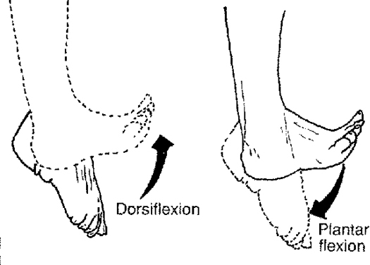
Plantar flexion describes the downward movement of the foot (calf raise). The strongest muscle group in your leg - the triceps surae - performs much of the plantar flexion. The triceps surae is composed of the by the soleus (deepest calf muscles) and the gastrocnemius (superficial double headed calf muscle). Both these muscles form the Achilles tendon, which then attaches to the posterior surface of your heel bone (calcaneus). When these muscles contract (or shorten) they pull your heel bone up, and push your toe down.

Pain, restrictions, tightness in these structures, or abnormal motion during plantar flexion is often the immediate cause of Achilles Tendonitis. In some cases, these same structures may also be affected by knee function and stability since the gastrocnemius muscle crosses three joints – knee, ankle, and subtalar joints. Alterations in the function of any of these joints will also have the immediate effect of increasing tension on the Achilles tendon.
 
Effects of Abnormal or Restricted Knee Flexion
Another common cause of Achilles Tendonitis are restrictions and injuries to the hamstrings. The hamstrings are the antagonists to the knee flexors. Restrictions in your hamstrings often prevents the full extension of your knee, resulting in increased tension in the Achilles tendon. Hamstring restrictions can create restrictions and tightness in the calf muscles, which in turn tightens the Achilles tendon.

Resolving Achilles Tendonitis
It is easy to see how a restriction in one area can cascade and develop into multiple restrictions within other associated structures. The key to resolving Achilles Tendonitis is to remove all these restrictions – along the entire kinetic chain – not just at the point of pain. Soft tissue therapy (ART, Graston, Registered Massage Therapy, Fascial Manipulation) can be very effective at doing this, but it requires someone who is trained in biomechanical analysis to locate all the affected areas.

Key Point: The location of these restrictions, or weak links, will vary from person to person. Each individual may present with the same diagnosis of Achilles Tendonitis, but the areas of restriction will often be completely different, and thus require completely different treatments.
In addition to removing and releasing these restrictions, it is important to use exercise to rehabilitate, strengthen, and restore the function of all the structures in this kinetic chain.


If you would like more information or to purchase our books please go to www.releaseyourbody.com . 

If you would like information about our clinic in Calgary Alberta please go to www.kinetichealth.ca.


(COPYRIGHT KINETIC HEALTH 2012 – ALL RIGHTS RESERVED)

Friday, February 3, 2012

Resolving Tension Headaches


Approximately 90 percent of headaches originate as tension headaches. Medical experts continue to debate over the causes of tension headaches. In my opinion, 90% of tension headaches are either caused by mechanical factors or are perpetuated by them. In other words, most ongoing, chronic, tension headaches can be attributed to specific physical restrictions. These are restrictions in muscles, ligaments, tendons, and fascia which cause nerve impingement syndromes, vascular changes, motion compensations and the output of biochemical substances that affect pain centres.

Symptoms of Tension Headaches
Tension headaches usually last from half-an-hour to an hour but they can continue to return for weeks, and in chronic cases, last for years. People who suffer from tension headaches often describe their headache as a dull ache, or they may experience them as a band of tightness around the sides of their head. This band may even feel like a vice compressing their skull. In severe cases the pain may even feel like a hooded cape that drapes over and across the shoulders. Obviously in such severe cases more than just the head is involved in creating the pain syndrome.
The following is a list of some of the common symptoms of tension headaches:
  • Band like pressure around the head.
  • Difficulty concentrating.
  • Difficulty sleeping (insomnia).
  • Fatigue and irritability.
  • Loss of appetite.
  • Neck, jaw/TMJ, or shoulder discomfort.
  • Severe pain behind the eyes.
  • Tenderness of the scalp.

You can usually differentiate a tension headache from other types of headaches because there are many symptoms you will not experience. For example, tension headaches do not cause visual disturbances, nausea, vomiting, numbness on one side the body, or slurred speech.

Triggers for Tension headaches
Tension headaches are triggered by numerous causes. Some of the potential triggers include:
  • Anxiety (including several medications used for reducing anxiety).
  • Arthritis
  • Clenching or grinding teeth (bruxism).
  • Dental Work.
  • Depression (including medications used for depression).
  • Holding one position for a long time, or working in awkward positions.
  • Inflammation of the neck or shoulders.
  • Lack of sleep or insomnia.
  • Lack of physical activity.
  • Overuse of headache medication (3rd most common cause of headaches).
  • Poor posture.
  • Stress.
  • Trauma.
  • Whiplash injuries (hyper-extension hyper-flexion injury)

Nerve Compression

Nerve compression can be a major factor in the initiation and perpetuation of tension headaches. If we look at the pathway of different peripheral nerves that transect, or pass under, musculoskeletal structures (muscles, ligaments, tendons, and fascia) it is easy to see how nerve compression can create a tension headache. Consider how a restriction of the following muscles can cause nerve compression which in turn can cause a tension headache.
Muscles:
o The Suboccipital nerve supplies input to the muscles of the suboccipital triangle. Compression of this nerve can occur at a muscle called the superior oblique.
o A trigger point in the superior oblique muscle itself will also refer pain to various regions of the head.
Referred pain from the trochlear region in tension-type headache: a myofascial trigger point from the superior oblique muscle. Headache 2005 Jun;45(6):731-7. Fernandez de las Peñas C, Cuadrado ML, Gerwin RD,Pareja JA.
· The greater occipital nerve is located directly under the semispinalis capitis muscle. Compression of this nerve is one of the causes of cervicogenic headaches. The symptoms from these headaches are called occipital neuralgias.
· Occipital neuralgia is a medical condition characterized by chronic pain in the upper neck, back of the head and behind the eyes. This is sometimes known as C2 neuralgia or Arnold’s neuralgia.
§ The lesser occipital nerve and the greater auricular nerve both pass by theSCM. Compression of these nerves will give symptoms of occipital neuralgia.
· The third occipital nerve travels under the trapezius muscle until it pierces this muscle and ends up in the lower part of the head (occiput). Compression of this nerve by a restriction in the trapezius muscle can also cause occipital neuralgias.

Biochemical Changes

If we review the literature on tension headaches there is no doubt that tension headaches are associated with changes in the levels of certain brain chemicals such as serotonin, bradykinins, and substance P.
  • Serotonin acts as a chemical messenger that transmits nerve signals between nerve cells. It also causes blood vessels to narrow.
  • Bradykinins mediates the inflammatory response, increases vasodilatation(expansion of arteries and veins), and causes contraction of smooth muscle.
  • Substance P functions as a neurotransmitter, especially in the transmission of pain impulses from peripheral receptors to the central nervous system.
The question is, are these chemical the initial factor that created the headache or are they a secondary consequence of physical restrictions. In my opinion, the physical changes probably took place first; which in turn caused a cascade of events that eventually increased, or altered, the levels of these chemicals.

Support for my perspective comes from what is called central sensitization. Central sensitizationhas been hypothesized as one of the primary mechanisms of chronic pain conditions. Central sensitization occurs when pain receptors in your nervous system (nociceptive neurons) become sensitized by tissue damage or inflammation.
By damage, I am not just referring to some type of trauma like a motor vehicle accident. I am also referring to damage caused by:
  • The micro trauma of repetitive strain injuries.
  • Stress caused by muscle imbalances.
  • Long-term stress placed on your body by poor posture.
  • De-conditioned muscles.
  • A thickening of the fascia. It is postulated that this thickening is caused by an increase in hyaluronic acid, which causes ‘densification’ of the fascia.
All these causes can produce areas of hyper-tonicity (increased tension) in our bodies that become chronically restricted; such as that huge knot most people have in their shoulders or the one just under the base of their skull.

When these hypertonic tissues becomes irritated or inflamed it releases chemical mediators (bradykinin, serotonin, substance P), which cause a sensitization of nerve endings resulting in pain, tension, and the production of headaches. If we review the history of most patients who suffer from tension headache we will routinely find a source of initial tissue damage (accident, trauma, history of tension) and associated inflammation which causes biochemical changes that elicit headaches.

Myofascial trigger points and sensitization: an updated pain model for tension-type headache Fernandez-de-las-Penas C, Cuadrado ML, Arendt-Nielsen L, Simons DG, Pareja JA. Cephalalgia. 2007 May;27(5):383-93. Epub 2007 Mar 14. inDepartment of Physical Therapy, Occupational Therapy, Physical Medicine and Rehabilitation, Universidad Rey Juan Carlos, Madrid, Spain.
Stress-induced pain and muscle activity in patients with migraine and tension-type headache. Leistad RB, Sand T, Westgaard RH, Nilsen KB, Stovner LJ. Cephalalgia. 2006 Jan;26(1):64-73. Department of Neuroscience, Norwegian University of Technology and Science and Department of Neurology and Clinical Neurophysiology, St Olavs Hospital, Trondheim, Norway.

Treatment of Tension Headaches

To resolve Tension Headaches it is essential that we remove all physical restrictions throughout the body’s Kinetic Chain. This would include restrictions in your shoulders, neck, jaw, skull, and perhaps other affected areas. In other words, wherever we find restrictions that may be initiating a physical cascade of events that will eventually cause physical or biochemical changes resulting in a headache. This will include:
It is extremely important to recognize that if we are only dealing with the headache on a pharmaceutical basis we have not addressed the chronic chain of events that perpetuates the headache.

There is a considerable amount of research in the literature to support this perspective. Even a change in posture can greatly affect the prevalence of a tension headache.
Research has demonstrated that tension or weakness in any of the following areas will cause an increase in both the intensity and frequency of tension headaches.
Examples of Anterior Structures:
Examples of Posterior Structures:
Our clinical experience has shown that the majority of chronic tension headaches can be completely resolved or substantially reduced by using soft tissue therapy (Active Release Techniques, Graston Techniuqe, Fascial Manipulation, or Massage therapy) in conjunction with the correct exercise program.
This information is derived from our Release Your Kinetic Chain Exercise Series. If you would like to more information or to purchase our books please go to www.releaseyourbody.com
If you would like information about our clinic in Calgary Alberta please go towww.kinetichealth.ca.
(COPYRIGHT KINETIC HEALTH 2012 – ALL RIGHTS RESERVED)

Sunday, January 29, 2012

Stress Fracture - What You Need To Know


Stress fractures are one of the most common, and potentially serious, overuse injuries. A stress fracture is an incomplete fracture that can occur anywhere in the body, and are typically caused by repetitive forceful actions. In contrast, most other types of fractures are caused by a single, direct, traumatic impact.

Stress fractures usually occur in weight-bearing bones, and are commonly seen in the metatarsals of the foot, bones of the ankle, shins (tibia), knees, and hips (the neck of the femur is one the worst stress fractures). Stress fractures are often caused by repetitive activities such as running, dancing, soccer, or any sport that involves high levels of repetitive actions. Stress fractures can eventually lead to a complete fracture.

How the injury occurs
Your body is continually remodeling your skeletal system based on the stresses that are placed upon it. This remodeling process gives your body the ability to handle increased loads without further injury. The problem arises in that your body can only adapt to these stresses at a certain rate, and there is a finite limit to the amount of stress to which it can adapt. Injuries to the bone (microscopic fractures) occur when increased stress is placed upon your body too quickly, or when too much load is applied.
In more technical terms, stress fractures occur in the early stage of bone remodeling whenosteoclastic reabsorbtion of bone, outstrips the osteoblastic development of new bone. This results in a weakened bone that is susceptible to injury. Osteoblasts and Osteoclasts control the amount of bone in your body:
· Osteoblast - Osteoblasts are cells which are responsible for bone formation. These cells produce osteoids, that are composed primarily of Type I collagen.
· OsteoclastOsteoclasts remove bone (reabsorb bone) by removing its mineralized matrix and breaking up organic bone.
Two Common Types of Stress Fractures
· Low-Risk Stress Fractures– An athlete can often heal these injuries by reducing activity, focusing on cross-training, nutritional supplementation, and a programs of conservative care accompanied by appropriate rehabilitation exercises.
· High-Risk Fractures – These fractures often occur on the tension side of a bone. Typically, these type of stress fractures do not respond well to any weight bearing activities or stresses, and require a non-weight bearing cast or complete immobilization to heal. In advanced cases surgery may be required to complete the healing process. Typical high risk areas include: Medial malleolus, navicular bone, fifth metatarsal, and the femoral neck.
You will need to consult a qualified medical practitioner to determine if a stress fracture is high-risk or not.

Risk Factors for Stress Fractures
Many factors can result in stress fractures including:
    • Faulty or worn-out equipment such as:
    • Training errors such as:
      • Sudden increases in frequency or duration of training.
      • Training while in a state of exhaustion.
      • Poor training techniques.
    • Amenorrhea or the absence of menstrual bleeding results in loss of bone mass at a similar rate to that experienced by post-menopausal women (up to 5% per year). This makes them very susceptible to stress fractures.
    • Restrictive diets that results in an inadequate supply of essential nutrients or calories, and can have a negative effect on bone density.
    • Muscular weakness or muscle imbalances.
Diagnosis
  • HistoryPatients who develop stress fractures often have a history of pain that is brought on by physical activity, and reduced pain with rest. There is usually an accompanying history of recent increases in physical activity.
  • Palpation – Touching the affected area (around the stress fracture) often elicits localized tenderness directly over the bone. Swelling over the area and redness is also commonly observed. Often there will be tenderness to percussion over the area.
  • Pain – Unfortunately, the pain pattern of a stress fracture may not provide a direct indication about the actual presence of a stress fracture. The pain from a stress fracture is usually well localized, but in some cases, can be spread out over a larger area.
      • Note: In the case of a femoral neck stress fracture (seen in runners/dancers or high impact athletes) the patient will feel groin pain that becomes worse with any activity (including walking) and that gets better with rest. This can become a very serious stress fracture if it transforms into a complete fracture. In such cases, the head of the femur can die due to lack of blood supply caused by the fracture.
  • Tuning Fork The vibrations of a tuning fork have been used as an indication of a stress fracture. Some practitioners dispute this test, but it may give the practitioner an indication that further diagnostic tests need to be performed.
  • X-rays – Diagnostic X-rays are often not very good at finding a recent stress fracture. A stress fracture may not show up on the X-ray for 2-3 weeks. Despite this, an X-ray is one of the first diagnostic procedures that should be performed. If possible an MRI, or bone scanshould also be performed, these procedures are much more definitive than an X-Ray.
Treatment Considerations: Keep the following points in mind when treating stress fractures.

What Not To Do for Stress Fractures
Certain treatments and behaviors will exacerbate, or inhibit the healing of your stress fractures, including:
  • DO NOT USE NASID’s – These medications inhibit bone healing and mask pain symptoms. Unfortunately many athletes, especially professional dancers, use large doses of NSAID’s.
  • Ignoring a stress fracture - A stress fracture that is ignored could turn into a complete fracture, which then requires surgery, resulting in a long healing time. Bottom line, you will never get over a stress fracture by just ignoring it, it will only get worse until even minor motion becomes extremely painful.
What To Do to Treat Stress Fractures
The following has been found to be effective in dealing effectively with stress fractures.
  • Prevention – The best way to treat this injury is to avoid it in the first place.
o Training No matter which sport or activity you are involved in (running, dance, etc.) be sure to increase your distance, frequency, or intensity gradually over a period of time.
§ Focus on the quality of your training, not just quantity. Adequate rest goes a long way in preventing a stress fracture. Rest is an essential training component for avoiding injury and stress fracture.
§ Do not train when you are exhausted, higher levels of injuries occur when in states of exhaustion.
§ Make Your Bones Strong (preferably before the injury occurs).
A key point here is that you can only make deposits of calcium (and other minerals) into your bone bank up until age 25. After age 25, you need to take daily calcium to prevent unnecessary withdrawals from your bones. Note of interest: Calcium deficiency is a prerequisite for a stress fracture. 98% of Adolecent dancers are calcium deficient (Harkness Centre for Dance Injuries 2010).
o First consider the composition of your bones:
§ 70% minerals (calcium phosphate, magnesium, sodium, potassium, fluoride, chloride). This bone matrix of mineral is called hydroxyapatite. You need all of these minerals. It pays to focus on good dietary habits including the right amount of supplementation.
o Maintain your calcium levels:
§ Children under 9 years of age require 800 mg. per day. If you are between 9 - 50 years old, you need 1300 mg of calcium per day. Those over age 50 require at least 1000 mg. of calcium per day.
§ 70% of your bones are made up of minerals (hydroxyapatite). The best way to ingest calcium is through a complete Bone Matrix supplements. We often recommend that our patients take MCHA to meet their nutritional requirements.

§ After the Stress Fracture has Occurred - The time frame for recovering from most lower-extremity stress fractures is between 8 to 17 weeks. This time frame will be much longer if the area is not given adequate time to heal. The good news is that most stress fractures respond well to conservative therapy and do not require surgery (if the stress fracture is not ignored).
o Initially, weight bearing exercises must be eliminated with any lower-extremity stress fracture. This is essential to allow the bone to heal and complete the normal remodeling process.
o An air cast (pneumatic walker/walker boot) or crutches is often recommended during the initial stages of injury. The air cast will often be used for 4 to 6 weeks depending on the speed of healing.
o Cross-training is essential to speed the recovery. Swimming, cycling, and routines that do not involve weight bearing are often recommended. The patient should also continue with upper extremity exercises.
o A slow rate of return to activity should be implemented since introducing weight-bearing exercises too soon will increase the duration of the injury. As a general rule, once the area is completely pain-free, light walking can be resumed.
o Pulsed electromagnetic field or low-intensity pulsed ultrasound therapy could speed the healing.
o Rehabilitation programs need to focus on several key factors (flexibility, strength, endurance, balance, propiroception, and alignment). This also includes addressing any weaknesses in hip or proximal stabilizers.
o Custom made orthotics may also be useful in helping to reduce biomechanical stresses on the affected area (Available through Kinetic Health).


(COPYRIGHT KINETIC HEALTH 2012 – ALL RIGHTS RESERVED)