Showing posts with label ankle injuries. Show all posts

Tuesday, February 7, 2012

Ankle Stability - The Retinaculum


Usually when we think about ankle problems, we think about sprained ankles or a strained muscle, not something called a retinaculum. Yet these fascial structures play a significant role in a wide variety of chronic ankle problems.

So what is a retinaculum? From one perspective a retinaculum is a band of thick deep fascia that holds the long tendons of your ankle (those that cross the ankle) in place. Retinaculum also acts as a pulley system increasing mechanical advantage.

From the second perspective retinaculum are a major source of neurological receptors involved in balance and proprioception. Essentially retinacula have been hypothesized as key structures in spatial control for foot and ankle movements.

The following section is an overview of specific retinacula and what structures pass underneath them. As you look over the individual sections of the retinaculum also think of these areas as part of one large fascial interconnecting unit.

Retinacula do not exist as they are illustrated
At the second international fascia conference in Amsterdam it became very clear to me that retinacula do not exist as they are illustrated in textbooks. There is a lot of interconnecting fascia that has to be removed before retinaculum look the way they are presented in text books. Research is now showing that these fascial connections (which are removed by dissection) are very important for both force transmission and neurological function.

Retinaculum Anatomy:
Front (Anterior) Ankle Retinaculum
Extensor retinaculum (2 parts)
o This structure holds in place tendons from the following muscles; tibialis anterior,extensor digitorum longus, extensor hallucis longus, and peroneus tertius.
o The deep peroneal nerve also passes under the retinaculum.
o The inferor retinaculum is shaped like a Y (once the entire surrounding fascia is removed) and has a lower and upper portion. The Y shape has the function of preventing “Bowstringing” of the tendons during ankle motion.

Pain/Symptom pattern: If there is a restriction in an extensor retinaculum, (front of the ankle) you may experience the following symptoms:
  • Localized pain or restriction on the front of ankle when running or walking. It is a very common symptom that I see with runners.
  • Tension can also alter the muscle firing patterns in the lower extremity. This can create a host of injuries and result in a substantial decrease in athletic performance
  • Outside (Lateral) Ankle Retinaculum
    Pain/Symptom pattern: Tension or a restriction in this area will often cause lateral ankle pain, altering both foot and ankle motion. This can easily lead to ongoing injury and a decrease in athletic performance.

    Note: Peroneal retinacula are often injured during ankle sprains (inversion injury). Anytime there is persistent pain after an ankle sprain, a retinaculum injury should be considered. For more information on ankle sprains read my six part blog on Ankle Sprains (Inversion Sprain).

    Inside (Medial) Ankle Retinaculum
    Pain/Symptom pattern:
    • Restrictions of the flexor retinaculum are associated with what is known as Tarsal Tunnel Syndrome. In this syndrome people experience sensation of: Pain, tingling and altered sensation anywhere from the ankle, heel, bottom of the feet, to the toes.
    • A person with this syndrome will often experience an electric shock sensation, which travels into the foot, when they tap directly over the retinaculum. This is also known asTinel’s sign.

    Tarsal Tunnel Syndrome (TTS): Tarsal Tunnel Syndrome refers to compression of the posterior tibial nerve in the flexor retinaculum.
    • Persons with flat feet (fallen arches) are susceptible to TTS. Also any type of enlargement in the Tarsal Tunnel can cause this syndrome which includes swollen tendon, cysts, arthritic bone spur, varicose veins, or even inflammation in the surrounding area.
    • If this syndrome is left to progress it can lead to permanent nerve damage.
    • Conventional therapy can often be very effective in treating this syndrome (Rest, Ice, and Exercise). Active Release Techniques has developed some specific protocols the release the posterior tibial nerve in the Tarsal Tunnel.

Injury To The Retinaculum
When functioning correctly, tendons glide under these retinacula without hindrance. With injury (trauma, repetitive strain), the retinaculum can become a site of tendon restriction, nerve impingement, and circulatory compression. Injury to the retinaculum will cause mechanical and neurological damage.
From a mechanical perspective, when tension is created between the retinaculum, and the structures that pass under them, a considerable amount of tension occurs. This tension can be a major problem since tension creates friction, which can cause micro-tears in the tissue,inflammation, and eventually adhesion formation. These adhesions inhibit relative motion, alter lower extremity biomechanics, and lead to a host of compensations.

From a neurological perspective, injury causes an alteration in neurological receptors (mechanoreceptors and proprioceptors). This leads to both ankle and foot instability. Instability of the ankle and foot creates abnormal motion patterns, compensations which can lead into numerous injuries throughout the body.

Treatment
Restrictions of the retinacula can be treated quite effectively with Manual Therapy (Active Release Techniques, Graston Technique, and Massage Therapy) and a series of corrective exercises. Treating with manual therapy involves breaking restrictions between the retinaculum and the tendon. Essentially the practitioner is restoring relative motion between the retinaculum and the tendons (and of course the muscles that the tendons are attached to).

The practitioners should also be focusing their treatment on the fascial lines of stress. Lines of stress in fascia are often created during injury in multiple locations not just at the site of pain. If these areas of fascial stress can be released, then normal fascial tension can be restored.
Restoring overall fascial tension, besides releasing adhesions between retinaculum and the soft tissues that pass under them, can have significant effects in resolving an injury.

Fascial interconnections are not theoretical entities; they are actual physical structures that have been mapped out. Researchers such as Thomas Meyers (Anatomy Trains) and Luigi, Carla, and Antonio Stecco (Fascial Manipulation) have spent decades researching these interconnections. During the second International Conference About Fascia at the University of Amsterdam, I had the privilege of listening to medical experts from around the world confirm this and related fascial research.
In my own clinical experience we have seen excellent results in improving ankle stability by removing adhesions at the retinaculum itself, but even better results when we work on restoring overall fascial tension

Exercise
Exercise plays a significant role in the rehabilitation of a retinaculum injury. Strengthening and flexibility exercises are needed, but because a significant component of a retinatculum injury involves neurological receptors, balance and proprioception exercises are also essential for full recovery.

The following links are examples of exercises that we often recommend for out patients with injuries to the retinaculum (from Core Performance).
If you would like more information or to purchase our books please go towww.releaseyourbody.com . 


If you would like information about our clinic in Calgary Alberta please go to www.kinetichealth.ca.

(COPYRIGHT KINETIC HEALTH 2012 – ALL RIGHTS RESERVED)

    Wednesday, February 1, 2012

    Medial Ankle Pain - Dancer’s Tendonitis


    An injury to the Flexor Hallucis Longus (FHL) tendon causes medial ankle pain or pain on the bottom of the foot. FHL injury is a condition that is often overlooked or misdiagnosed. This injury affects dancers, runners, soccer players, and any other athlete who performs repeated, propulsive forces, or jumping. Injury of theFlexor Halicus Longus
    muscle is sometimes called “Dancer’s tendonitis” but it is not limited to just dancers. Image
    Alvin Ailey American Dance Theater dancer Constance Stamatiou.


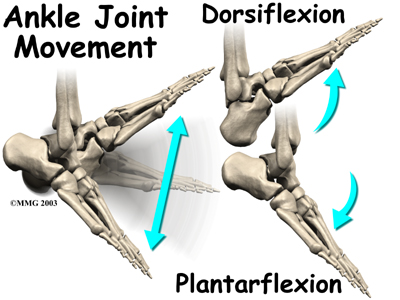
    The Flexor Hallucis Longus (FHL) muscle allows you to point your big toe (plantar-flexing your big toe) and stabilizes the Subtaler Joint. The Subtaler Joint is located between two bones in your ankle - the Talusand the Calcaneus. The Subtaler Joint allows movement of the heel toward the medial plane (inversion) as well as movement of the heel towards the lateral plane (eversion).

    Anatomy/Biomechanics of Medial Ankle Pain
    The Flexor Hallucis Longus muscle is located deep under your calf muscles (the most lateral muscle of the deep compartment). The FHL originates on the lateral lower leg (distal 2/3 of Fibula). It then travels at an oblique angle (crosses the posterior surface of Tibia) down towards the medial ankle (posterior surface of Talus) and travels under a section of the heel bone (Sustentaculum Tali of Calcaneus). The FHL then passes under the sole of the foot (between the two heads of the FHB) and inserts into the base of the big toe (base of the Distal Phalanx of Hallax).

    As mentioned the FHL Tendon curves around a structure called the Sustentaculum Tali. TheSustentaculum Tali is a bony section on the inside of the heel bone (Calcaneus). This is often a site of friction and irritation for the FHL tendon. The FHL tendon also travels between the twoSesamoid bones just behind the big toe (Metatarso-Phalangeal Joint). The sheath that surrounds the FHL tendon is often irritated.

    Causes and Presentation of Medial Ankle Pain
    The repetitive motion of pushing-off with your foot (plantar flexion) during dance, running, soccer, and jumping can cause injury to the FHL.

    Injury to the FHL tendon and muscle can present in a variety of ways - sometimes involving inflammation and sometimes not. Injury to the tendon without inflammation is refer to as a “Tendinopathy”. Tendinopathy refers to a degenerative lesion in the tendon without affecting the tendon sheath that surrounds the tendon.

    When FHL inflammation (tendonitis) is present in the foot, it usually occurs in one of the following three locations.
    2. At the “Knot of Henry” – a section just behind the big toe (first metatarsal) where the FDL muscle crosses the FHL tendon.
    3. Just behind the big toe by the Sesamoids bone.
    o A Sesamoid bone holds its tendon away from the center of the joint and acts to increase mechanical force.

    When the FHL tendon becomes nodular, a condition called Hallux Saltans can develop. Hallux Saltans is similar to trigger finger in the hand, except it occurs in the big toe. Triggering of the toe occurs when the nodular thickening of the tendon snaps through the fibro-osseous tunnel. This causes a jerking motion, much like a trigger finger.

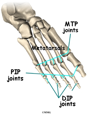
    If not addressed Hallux Saltans can contribute to the progression of an additional condition called Hallux Rigidus. Hallux Rigidus means "stiff great toe". Hallux Rigidus is the second most common disorder of the first MTP joint. The most common injury is a bunion - otherwise known as Hallux Valgus.


    Special Considerations For The Dancer
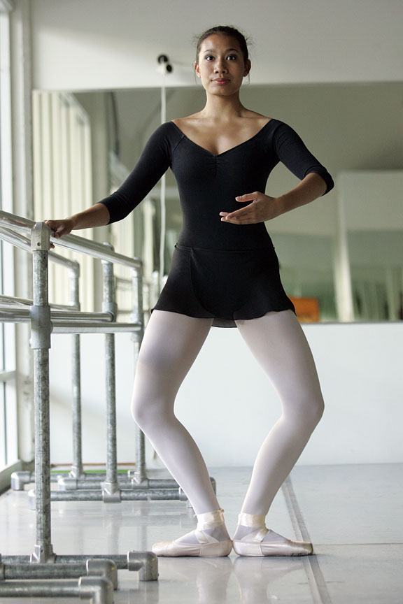
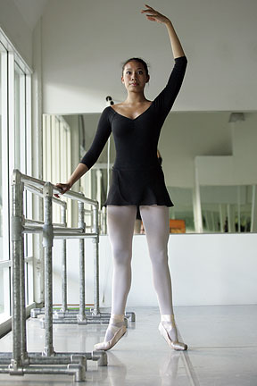
    Injury to the FHL in dancers is often caused by the repetitive motion of changing position from a pliĂ© position to a relevĂ© position. This action produces a force that is 10 times the dancer’s body weight.

    Ideally, for greater stability and increased propulsion, the foot should be in a supinated position at the heel during push-off. For a dancer, any action that causes a reduction in plantar flexionmotion can create an FHL injury. Biomechanically, a lack of plantar flexion leads to a prolonged pronation position of the foot when pushing off in the Propulsion Phase.
    In dancers, the FHL tendon is often compressed while performing a relevé position and is over-stretched while performing a plié postion. In such a case, the dancer will feel posterior medial ankle pain when performing the plié. (Photo - Dance Theatre of Harlem).

    Diagnosis and Imaging - FHL Injury
    X-rays for this condition are not very specific. X-Rays are good for ruling out fractures (Calcaneus, Distal Medial Malleolus, or Os Trigonum) that may cause an impingement of the FHL tendon. It is important to note that X-Rays will not provide a definitive diagnosis of FHL injuries.
    A good history is much more definitive, especially when accompanied by a full physical examination and an MRI. MRI imaging is an excellent resource for showing damage to tissue fibers, inflammation, and swelling (edema).
    If the MRI is not available or is too expensive, a CT scan (Computed Tomography) can prove to be a useful alternative.

    Treatment
    In most cases, FHL syndrome (involving partial tears, tendinosis, or inflammatory conditions) responds well to conservative care. However, a complete tear of the FHL tendon may require corrective surgery since serious cases of FHL injury could end an athlete’s career.

    Conservative care includes:
    · Ice or heat depending on stage and type of injury (inflammation or not). See our Icing andHeating blog.
    · Reduction in activity. Usually, some modification in all activities is necessary.
    · Supports (crutches or walker boot) can be beneficial.
    · Taping – Kinesio Taping. At Kinetic Health we use SpiderTech Kineso-taping.
    · Soft-tissue and joint mobilization (Active Release, Graston Techniques, Massage, and Fascial Manipulation).
    · Inflammatory strategy implementation (medication for no longer than seven days). See ourblog about Reducing Inflammation.
    · Exercises for increased strength, flexibility and balance. Go to www.releaseyourbody.comfor exercises you can use to treat this condition.
    · Biomechanical corrections (footwear or Orthotics) .
    · A gradual return to activities.

    If you would like more information or to purchase our books please go to www.releaseyourbody.com . 

    If you would like information about our clinic in Calgary Alberta please go to www.kinetichealth.ca.


    (COPYRIGHT KINETIC HEALTH 2012 – ALL RIGHTS RESERVED)


    Wednesday, September 8, 2010

    Ankle Sprain (Inversion Sprain) – Part 6


    Restoring Flexibility to the Ankle
    It is extremely important to stretch after experiencing an ankle injury of any kind. During the recovery phase, your body forms and lays down collagen to repair the injured area from two days, and up to 6 weeks after, the injury occurs.
    If you suffer from an inversion sprain, and perform the correct stretching exercises, then you can ensure that the majority of the collagen that is being laid down, is being laid in the same direction as the tissue which is being repaired. This makes the repaired tissue stronger and more capable of performing its function in the future. However, if you do not stretch the injured area, the new collagen will be laid down in more random patterns, leading to the development of weaker tissue (scar tissue) that is easily re-injured

    Here are some of the areas for which we recommend stretching exercises when our patients are recovering from an ankle injury:
    • Calf muscle (gastrocnnemius and soleus) - Stretch the calf muscle as it often becomes very tight after an ankle sprain, as these structures try to protect the injured joint.
    • Peroneal muscles – Stretch the peroneals (along the sides of your calf) as these muscles are often injured along with the ligaments in the ankles.
    • Iliotibial Band – Yes, these tissues are in your upper leg, but there are direct fascial connections from the peroneal muscles into the IT band. A injury to the peroneal muscle may cause compensation injuries up into the IT Band. So be sure to perform stretches to release the IT Band.
    • Gluteal muscles – Since the IT band is formed from the deltoid complex (gluteus maximus, gluteus medius, and tensor fascia latae, problems in the IT band will affect hip function. So it is important to release the muscles of the gluteals with stretches and foam rollers.


    Proprioceptive Training

    Whenever an inversion sprain occurs, the injury is usually not restricted to just the ligaments, tendons, muscle fibers, and fascial fibers. Often, the embedded neurological structures within these soft-tissue structures are also damaged. These neurological structures (golgi tendon organs, muscle spindles, and joint receptors) perform an essential role in positional control. Any damage to these structures can have the effect of decreasing stability, which can lead to chronic ankle problems.
    Fortunately this damage can be repaired with exercise protocols that involve balance and proprioception. Some of the proprioceptive exercises we use with our patients at the clinic are:
    • One-legged Stand
    This is a good initial exercise to try, with a slow progression into partial single leg squats. Do all of this exercise within a pain free range-of-motion.
    • Wobble board training
    Slowly increase the difficulty of wobble board exercises from the two-legged balance exercises, into a single-leg exercise that combines full-body motions. Once you are ready, you can even try the single-legged version with your eyes closed. Note: The eyes-closed version should only be attempted after you are fully recovered and you are ready to work on increasing athletic performance.
    · Upside Down Bosu Ball
    The Bosu Ball is a great device for dynamically stretching and strengthening the ankle. Try this Side-to-Side Peroneal Stretch.
    § Turn the Bosu Ball upside down, balance on the flat surface, with your feet about shoulder-width apart.
    § Shift your weight to the right side so that the right edge of the Bosu Ball almost touches the ground. Then immediately shift your weight to the left side so the left edge of the Bosu ball almost touches the ground. Continue to shift from side-to-side. As you do this exercise, you should feel a stretch in the muscles along the lower outside of your leg (peroneals).
    § After a few minutes of performing this action your lower extremity muscles should start to feel fatigued.
    § Once you become comfortable with balancing the side-to-side actions, you can start integrating arm actions into this exercise. As you shift your lower extremity to the right side, swing both your arms to the left. When you shift your weight to the left side, swing your arms to the right. This simulates a downhill skiing (cross crawl) type of action that integrates all the muscles of your body.

    Sport Specific
    If you are involved in a particular sport, take the time to create a balance/proprioceptive exercise that matches your specific sport. For example:
    · If you are a soccer player who is rehabilitating a sprained ankle, you may want to try one-legged stand on the injured ankle, while kicking a soccer ball with the other foot. To increase the level of difficulty, try standing on a balance pad with the injured leg, and then kick the ball with the other.
    · If you are a dancer suffering from an inversion sprain, try moving through your basic dance positions while on a wobble board or balance pad.
    · If you are a basketball player, try standing on a wobble board with the injured leg and practice throwing hoops.
    Be creative with your individual sport. Use wobble boards, balance pads, half-foam rollers, or bosu balls to rehabilitate the damage to your nervous system that is caused by an inversion sprain.

    Bottom line - inversion sprains are a common, yet complex, injury. This injury can involve multiple structures across numerous joints, and may even extend a considerable distance up the body. To correctly rehabilitate the injury, you must use an approach that takes into consideration musculoskeletal connections, the nervous system, and appropriate tissue remodelling.

    If you would like more information or to purchase our books please go towww.releaseyourbody.com . 

    If you would like information about our clinic in Calgary Alberta please go to www.kinetichealth.ca.


    (COPYRIGHT KINETIC HEALTH 2012 – ALL RIGHTS RESERVED)