Showing posts with label ankle exercises. Show all posts

Wednesday, September 8, 2010

Ankle Sprain (Inversion Sprain) – Part 6


Restoring Flexibility to the Ankle
It is extremely important to stretch after experiencing an ankle injury of any kind. During the recovery phase, your body forms and lays down collagen to repair the injured area from two days, and up to 6 weeks after, the injury occurs.
If you suffer from an inversion sprain, and perform the correct stretching exercises, then you can ensure that the majority of the collagen that is being laid down, is being laid in the same direction as the tissue which is being repaired. This makes the repaired tissue stronger and more capable of performing its function in the future. However, if you do not stretch the injured area, the new collagen will be laid down in more random patterns, leading to the development of weaker tissue (scar tissue) that is easily re-injured

Here are some of the areas for which we recommend stretching exercises when our patients are recovering from an ankle injury:
• Calf muscle (gastrocnnemius and soleus) - Stretch the calf muscle as it often becomes very tight after an ankle sprain, as these structures try to protect the injured joint.
• Peroneal muscles – Stretch the peroneals (along the sides of your calf) as these muscles are often injured along with the ligaments in the ankles.
• Iliotibial Band – Yes, these tissues are in your upper leg, but there are direct fascial connections from the peroneal muscles into the IT band. A injury to the peroneal muscle may cause compensation injuries up into the IT Band. So be sure to perform stretches to release the IT Band.
• Gluteal muscles – Since the IT band is formed from the deltoid complex (gluteus maximus, gluteus medius, and tensor fascia latae, problems in the IT band will affect hip function. So it is important to release the muscles of the gluteals with stretches and foam rollers.


Proprioceptive Training

Whenever an inversion sprain occurs, the injury is usually not restricted to just the ligaments, tendons, muscle fibers, and fascial fibers. Often, the embedded neurological structures within these soft-tissue structures are also damaged. These neurological structures (golgi tendon organs, muscle spindles, and joint receptors) perform an essential role in positional control. Any damage to these structures can have the effect of decreasing stability, which can lead to chronic ankle problems.
Fortunately this damage can be repaired with exercise protocols that involve balance and proprioception. Some of the proprioceptive exercises we use with our patients at the clinic are:
• One-legged Stand
This is a good initial exercise to try, with a slow progression into partial single leg squats. Do all of this exercise within a pain free range-of-motion.
• Wobble board training
Slowly increase the difficulty of wobble board exercises from the two-legged balance exercises, into a single-leg exercise that combines full-body motions. Once you are ready, you can even try the single-legged version with your eyes closed. Note: The eyes-closed version should only be attempted after you are fully recovered and you are ready to work on increasing athletic performance.
· Upside Down Bosu Ball
The Bosu Ball is a great device for dynamically stretching and strengthening the ankle. Try this Side-to-Side Peroneal Stretch.
§ Turn the Bosu Ball upside down, balance on the flat surface, with your feet about shoulder-width apart.
§ Shift your weight to the right side so that the right edge of the Bosu Ball almost touches the ground. Then immediately shift your weight to the left side so the left edge of the Bosu ball almost touches the ground. Continue to shift from side-to-side. As you do this exercise, you should feel a stretch in the muscles along the lower outside of your leg (peroneals).
§ After a few minutes of performing this action your lower extremity muscles should start to feel fatigued.
§ Once you become comfortable with balancing the side-to-side actions, you can start integrating arm actions into this exercise. As you shift your lower extremity to the right side, swing both your arms to the left. When you shift your weight to the left side, swing your arms to the right. This simulates a downhill skiing (cross crawl) type of action that integrates all the muscles of your body.

Sport Specific
If you are involved in a particular sport, take the time to create a balance/proprioceptive exercise that matches your specific sport. For example:
· If you are a soccer player who is rehabilitating a sprained ankle, you may want to try one-legged stand on the injured ankle, while kicking a soccer ball with the other foot. To increase the level of difficulty, try standing on a balance pad with the injured leg, and then kick the ball with the other.
· If you are a dancer suffering from an inversion sprain, try moving through your basic dance positions while on a wobble board or balance pad.
· If you are a basketball player, try standing on a wobble board with the injured leg and practice throwing hoops.
Be creative with your individual sport. Use wobble boards, balance pads, half-foam rollers, or bosu balls to rehabilitate the damage to your nervous system that is caused by an inversion sprain.

Bottom line - inversion sprains are a common, yet complex, injury. This injury can involve multiple structures across numerous joints, and may even extend a considerable distance up the body. To correctly rehabilitate the injury, you must use an approach that takes into consideration musculoskeletal connections, the nervous system, and appropriate tissue remodelling.

If you would like more information or to purchase our books please go towww.releaseyourbody.com . 

If you would like information about our clinic in Calgary Alberta please go to www.kinetichealth.ca.


(COPYRIGHT KINETIC HEALTH 2012 – ALL RIGHTS RESERVED)

Tuesday, September 7, 2010

Ankle Sprain (Inversion Sprain) – Part 5


Exercise Rehabilitation For Ankle Sprains
When prescribing ankle exercises, we must take into consideration the multiple structures that are involved in performing and coordinating ankle motion. To attain a full resolution from an inversion sprain, we must be sure to consider multiple muscles and complex fascial layers, all of which work together across numerous joints.
To do this, we must be sure to address three essential components: strength, flexibility, and proprioception.
Strengthening the Ankle

Strengthening exercises are essential! Manual therapy, by itself, will not fully prevent an injury from returning without the addition of strengthening exercises. This is because the type and quality of tissue remodeling that occurs after an injury is dependant on the type of forces that are applied to that tissue. When appropriate strengthening exercises are applied, the collagen remodels to withstand the stresses that are placed upon it. The possibility of re-injury is very high without appropriate strength training.

Strengthening exercises for an inversion sprain can be subdivided into several categories depending on the stage of the injury. The following subcategories describe stages from non-weight-bearing to full-weight-bearing. Note: These are examples of possible recommendations, but actual exercise recommendations will vary from case to case.

1. Active Range Of Motion (AROM) – Non-weight bearing exercises
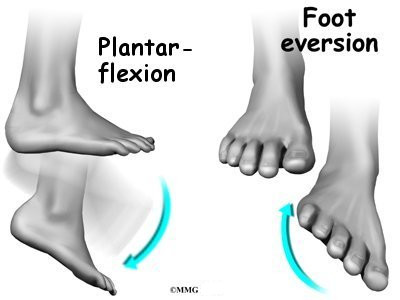
a. Four basic foot motions – Perform the following four basic motions: plantar flexion, dorsi flexion, inversion, and eversion. Perform these foot motions slowly, holding the end range of each position for 5-10 seconds. Repeat the action several times.
b. Alphabet exercise – Use your foot to draw the letters of the alphabet in the air, allowing your ankles to rotate with each motion

2. Isometric strengthening of the ankle – Isometric exercises are performed in static positions (unlike dynamic exercises which are performed through a range of motion).
a. The Isometric Four – In this exercise, you need to resist each basic motion (plantar flexion, dorsi flexion, inversion, and eversion) of the ankle. For example:
· Start with your ankle in neutral position, with your foot braced against a wall or table.
· Then, without allowing your foot or ankle to move, try to perform each of the four basic actions. This causes all the muscles involved in the action to contract.
· Hold this contraction for 5-10 seconds and relax for about 10 seconds.
· Repeat this process for 4-8 repetitions, slowly increasing the number of repetitions.
· Repeat this routine for each of the four basic actions.

3. Resisted Strengthening of the Ankle – using Theraband or Tubing
a. Four-Way Resisted Tubing – In this series of exercises, we recommend using a theraband or tubing as you perform each of the four basic ankle motions. For example, with the inversion action:
· Attach a theraband around your ankle and attach the other end to a table or door.
· Against resistance, roll your foot inward (invert your ankle) and hold for 3 to 5 seconds.
· Repeat for 10 to 15 repetitions, increasing the number of sets from 1 set/day to 4 sets/day as you increase your ankle strength.
· Repeat this routine for each of the four basic ankle actions.

4. Partial Weight Bearing Ankle Exercise
a. Shifting Your Weight – In this exercise, lean against a counter or table, and try shifting some of your weight onto the injured leg. Start with just a few seconds of weight, and then increase this time, as the ankle permits. Repeat this exercise about 10 times. Be careful not to do too much, too soon.

5. Full Weight Bearing Ankle Exercises
Once the injured ankle is able to bear the full weight of your body, there are many exercises that will help to further strengthen the ankle. One of my favorites is the Eccentric Calf Raise.
Eccentric Calf Raises - One Legged
(Actually…these are calf drops, not raises)
In this exercise, the rising up portion is not that important. The key lies in the slow lowering of the body, as this builds strength without causing further injury to the body.
· Stand on the edge of step or curb, heels hanging over the edge of the step, and rise up on both legs in a classic calf raise.
· Now, using only one leg, slowly drop down to the starting position for a count of three
· Do 5 to 15 repetitions in each set. Start with two sets and gradually increase to 4 sets as your ankle strength increases.
· Allow your ankle to build up its strength slowly. Do not increase your repetitions and sets until you can complete each repetition with perfect form, and without pain.

Once you are able to properly perform full weight bearing exercises, you should consider beginning a routine that exercises your full body. These exercises should activate the knee, hip, and core.


In Part Six of Ankle Sprains we will cover stretching and proprioceptive exercises for resolving ankle sprains.


If you would like more information or to purchase our books please go towww.releaseyourbody.com . 

If you would like information about our clinic in Calgary Alberta please go to www.kinetichealth.ca.


(COPYRIGHT KINETIC HEALTH 2012 – ALL RIGHTS RESERVED)


Friday, September 3, 2010

Ankle Sprains or Inversion Sprains – Part 4


Fascia and Inversion Sprains
Fascia was once thought to be nothing more than packaging material covering muscles and organs. Back in school, when we worked in the dissection lab, we went out of our way to remove this material in order to expose those structures that were considered to be more important (an experience that I have heard repeated by many individuals in the medical field). Unfortunately, this lead to the very misleading perspective that fascia was unimportant.

Now, 20 years later I, and many others, have come to a very different conclusion. Fascia is everywhere, weaving through, and interconnecting every component of our body. Robert Schleip from Ulm University in Germany (of the Fascia Research Project) describes fascia extremely well when he describes fascia as “the dense irregular connective tissue that surrounds and connects every muscle, even the tiniest myofibril, and every single organ of the body forming continuity throughout the body.” In other words, fascia is the physical manifestation of the kinetic web that we have discussed so many times in our blogs and books.

Schleip R. Fascial plasticity—a new neurobiological explanation: part 1. J Bodyw Mov Ther. 2003;7(1):11–19.

Fascial interconnections are not theoretical entities; they are actual physical structures that have been well mapped out. Researchers such as Thomas Meyers (Anatomy Trains) and Luigi, Carla, and Antonio Stecco (Fascial Manipulation) have spent decades researching these interconnections. During the last International Fascia Research Congress at the University of Amsterdam, I had the privilege of listening to medical experts from around the world confirm this and related fascial research. (Take a look at these excellent articles for more details.)

Fascia as an Anchor for Force Generation

When one considers the prevalence of fascia throughout the body, it is not surprising to discover that it actually plays a critical role in all bodily actions, and that it is much more than the “bubble wrap” that many considered it to be.
In conventional medical theories, muscles have both an origin and insertion at the bone(s). In this view, when a muscle contracts, it pulls the origin toward the point of insertion to create motion. In reality, muscle fibers actually originate from, and insert into, fascial fibers. These fascial fibers, in turn, insert into multiple regions of the bone, and even into adjacent muscles. These additional points of contact and control provide the muscle with the ability to generate force in multiple directions.
In reality, when a muscle contracts to perform an action, only certain sections of the muscle contract. Force is not generated from just the origin and insertion points, but also through the numerous fascial connections. Anatomists at the University Maastricht in the Netherlands have done some incredible research that confirms this understanding.

Van Mameren H. Reaction forces in a model of the human elbow joint. Anat Anz. 1983;152:327–328.

Fascia as a Neurological Feed Back Loop

Muscles work together as functional units that coordinate their actions across multiple joints. Depending on the degree of motion required, and amount of force that is needed, each muscle will contract only specific areas of the muscle, rather than the entire muscle. As incredible as it may sound, these very specific motions are largely coordinated by the neurological receptors embedded in the fascia, and not by the brain. This is a form of non-localized intelligence, a concept that has long been postulated, but is only now being acknowledged as occurring in all life forms.

Fascia is full of two types neurological receptors - mechanoreceptors and proprioceptors. A mechanoreceptor is a sensory receptor that responds to mechanical pressure or distortion. A proprioceptor is a sensor that provides the brain with information about joint angle, muscle length, and tension.
Motion is coordinated by the feedback of the neurological receptors in the fascia when tension is transfered from one area to the next. (Previously these receptors were only thought to be located within just the muscles).

Fascial Lines and an Inversion Sprain

When we evaluate an inversion sprain of the ankle, we must also take into consideration all the fascial connections to the structures that are involved in performing and coordinating ankle motion. It is important to look at the big picture, and take into consideration that multiple muscles, working across multiple joints (all of which are interconnected by fascia) are all required to coordinate these motions.

In the case of an inversion sprain, the fascial connections we should consider is in the Lateral Line (see Anatomy Trains by Thomas Meyers). The following is a short synopsis of the lateral line. I have also included a link to the Anatomy Trains dissection video about the Lateral Line – Thomas Meyers Lateral line Video.

Lateral Line
· Beginning at the foot with the peroneal muscles, fascial interconnections travel up the outer leg to just below the knee (fibular head). This fascia then connects directly into the lower IT band into the deltoid complex (gluteus maximus, gluteus medius, and tensor fascia latae).
· The fascia then connects to the pelvis (iliac crest), which connects into the abdominal muscles (internal and external obliques), and then into the quadratus lumborum which connects into the ribs and the spine.
· The fascia then travels up between the ribs (intercostals muscles) and continues up the body to connect into the fascia of the neck (SCM, splenicus cervicis, and scalenes).

Once you see these videos, you will find it very hard to dispute the importance of these fascial connections.

Bottom Line: You can try treating inversion sprains with all the right techniques (ART, Graston, Massage etc.) and find that you still have not resolved a chronic inversion sprain if you did not consider, and treat, the fascial interconnections

Manipulation

In addition to the soft-tissue treatment component, you must also address the osseous (bone/joint) component. Joint manipulation helps to restore active range of motion in the ankle and speed recovery. It is very common for several of the joints of the feet to become stiff and rigid, due to either the initial injury or due to soft-tissue compensations over a period of time.

The exact area that requires joint manipulation varies depending on the severity of the sprain, number of reoccurrences, and the length of time that has passed since the injury occurred. The longer you wait, the more compensations your body must make, and the longer it will take to treat and recover from the injury.

In part five of Ankle Sprains we will provide Rehabilitative Exercises For Ankle Sprains.


If you would like more information or to purchase our books please go towww.releaseyourbody.com . 

If you would like information about our clinic in Calgary Alberta please go to www.kinetichealth.ca.


(COPYRIGHT KINETIC HEALTH 2012 – ALL RIGHTS RESERVED)

Thursday, September 2, 2010

Ankle Sprains or Inversion Sprains – Part 3


Scar Tissue
So, why is scar tissue formation such an issue when resolving ankle sprains? When ligament fibers are torn, the body lays down collagen in an attempt to support, reconnect, and reinforce the damaged tissue. This collagen is what we commonly refer to as scar tissue. Scar tissue is much weaker, less flexible, and easier to tear than normal ligaments. Scar tissue is weak and inflexible because it is laid down in a random pattern and does not follow the same fiber orientation as the normal ligaments. These areas of scar tissue formation have a different consistency and strength than the normal ligaments. (Image Body Worlds - permission granted).

This alteration in ligament consistency then creates instability in the ankle, resulting in abnormal motion patterns, which then can result in further injuries. A considerable amount of scar tissue can form with repeated ankle sprains (which are common). Although the initial intention of the body was to stabilize the ankle with scar tissue, it can have the effect of de-stabilizing it instead, due to the random patterns of fiber orientation.
Therefore, it is essential to remove this scar tissue, thereby helping in the restoration of normal motion patterns, and increasing flexibility and strength.

Manual Therapy for an Ankle Sprain

Manual therapy can make a huge difference in the recovery and prevention of an ankle sprain. By manual therapy, I am referring to techniques such as Active Release, Graston Technique, Massage Therapy, Fascial Manipulation, Manual Manipulation, and other manual procedures. In my opinion, these procedures are essential in the rehabilitation of an ankle sprain since they all act to break down and prevent scar tissue formation.

Manual therapy also speeds healing by increasing blood supply, oxygen, essential nutrients, and displace waste products that accumulate after an injury. This is especially important in treating ligaments because they generally have a very poor blood supply to begin with.

It is Important to Treat More Than Just the Ligaments

Earlier, I mentioned that there might be damage to several types of structures after an ankle sprain, especially the connective tissue, tendons, muscles, and nerves. The following is list of structures that are often injured in a lateral ankle sprain (inversion sprain).

Ligaments
• Anterior talofibular ligament (ATFL) – Most common injured structure.
• Calcaneofibular ligament (CFL) – Second most commonly injured structure.
Tendons
o With an inversion sprain, it is common for the tendons of the peroneal muscles to be injured. It also common for the peroneal tendon to move out of its normal position (subluxate) during the recovery stage of an ankle sprain.
Muscles
• Peroneus brevis muscle – Longitudinal tears of the peroneus brevis are commonly associated with lateral ankle sprains.
• Peroneus longus muscle – These muscles often show a different activation pattern when there is ankle instability. This is often due to restrictions that have formed in the muscle. These restrictions, if not removed, could make a person more susceptible to future injuries.
Both muscles, evert the foot at the tarsal joint, and plantarflex the foot at the ankle.
Retinaculum
o During a lateral ankle sprain, the foot rolls inwards with considerable force. This forceful action can tear the peroneal retinaculum. This retinaculum is a band of connective tissue that keeps the peroneal tendons in place. When a tear in the retinaculum occurs, patients will notice a snapping sensation in the lateral ankle. Conservative treatment is 4 to 6 weeks in a short leg cast. However, a severe tear of the retinaculum is not a candidate for standard manual therapy and may require corrective surgery.
Nerves
• Superficial peroneal nerve - This nerve is at risk for traction injury during a lateral ankle sprain (inversion sprain).

In Part Four of Ankle Sprains we will cover Fascia and Inversion Sprains as well as the Benefits of Joint Manipulation in the treatment of ankle sprains.


If you would like more information or to purchase our books please go to www.releaseyourbody.com . 

If you would like information about our clinic in Calgary Alberta please go to www.kinetichealth.ca.


(COPYRIGHT KINETIC HEALTH 2012 – ALL RIGHTS RESERVED)