Showing posts with label knee injury treatment. Show all posts

Sunday, March 20, 2011

Treating and Preventing Meniscus Injuries - Part 5


Exercise
If the injury is not severe enough to warrant surgery therapy can usually begin almost immediately. If surgery is warranted, exercises can usually begin within a short period of time [depending on your surgeon’s advice].
Initially, patients with meniscus injury tend to do better with non-weight-bearing activities that do not cause compression. Especially in cases of more severe injury it is important to progress to weight-bearing functional activities.
If you’ve had a posterior meniscus injury you should keep your knees ranges of motion limited while performing exercise. This will help to ensure that further injury does not take place. It is also important to avoid any rotational movements of the knee, especially in weight-bearing positions.
In cases of surgery it may be necessary to avoid any type of flexion exercise for up to two months. Exercises that include twisting activities or squats may need to be avoided for up to 3 to 6 months. Depending upon the degree of injury it may also be necessary to limit athletic activity for 4 to 6 months after surgery.
The following exercise recommendations are only general in nature for individuals who do not need surgery, or have been cleared by their surgeon to perform these exercises.
Initial Phase of Exercise
The objectives during this phase are to decrease swelling [RICE], prevent further injury [bracing may be necessary], prevent muscle atrophy and maintain overall strength. During this initial phase which is can be several weeks depending on the degree of injury, four weeks if post operative, further injury must be prevented. During this stage appropriate exercises would include:
  1. Isometric exercises for the Quadriceps (especially the VMO)
  2. Range of motion exercises [passive and active if appropriate]
  3. Stationary cycling - single leg cycling only using the uninjured leg
Intermediate stage of exercise (Patient can now flex and extend knee)
The objective of this stage is to return to full weight bearing on the injured leg and to increase muscular endurance. It is important to combine soft tissue mobilization with exercise to avoid scar tissue formation. During this stage appropriate exercises would include opened chain kinetic exercises such as:
  1. Limited open chain resisted tubing exercises of the ankles knee and hip.
  2. Stationary cycling with no or only minimal tension.
  3. One leg stands - in this exercise you alternate standing on one leg for approximately 20 to 30 seconds, shifting your weight back and forth. This can only be done if you are able to tolerate weight-bearing.
  4. An upper extremity weight training program
  5. Deep water running program
  6. Stretches should include the: Quadriceps, hamstrings, hip rotators, iliotibial band, and calf muscles.
As the patient progresses Open Kinetic and Closed Kinetic Chain resisted exercises can be used.
Advanced stage of exercise (Patient now has full range of motion)
  1. Begin running at 50 to 60% of effort.
  2. Step ups, onto box.
  3. Jumping and hopping exercises. These exercises must be performed completely pain-free.
  4. Eventually exercises should simulate activities performed by the sport or activity the individual wishes to return to,
  5. Balance and stability training on a gym ball can be used at this stage.
Conclusion:
In many cases meniscus injuries can be prevented. Active Release Techniques has been proven to be a viable option in removing meniscus entrapment’s before the actual tear occurs. Once an actual injury has occurred manual therapy and exercise should be implemented as soon as possible. Therapy must always consider key kinetic chain relationships to prevent an ongoing cycle of injuries. In addition to therapy exercise is essential without the correct rehabilitative strategy the probability of ongoing injury is almost 100%.
If you would like more information or to purchase our books please go to www.releaseyourbody.com . 

If you would like information about our clinic in Calgary Alberta please go to www.kinetichealth.ca.

(COPYRIGHT KINETIC HEALTH 2012 – ALL RIGHTS RESERVED)


Thursday, January 27, 2011

Treating and Preventing Meniscus Injuries - Part 2


In our previous blog (link) we discussed the causes and structural components of the knee and its menisci. Now…lets look at how we can treat injuries to the menisci.


Degree of Injury
Injuries to the menisci can range from minor, moderate, to more severe. Pain and swelling are the most common symptoms of a meniscus tear. With more severe menisci injuries, a person may notice some degree of “locking of the joint” accompanied by considerable instability of the knee. If “locking of the joint” occurs, it is imperative to immediately obtain medical attention from a sports physician, or some other medical practitioner who works with physical medicine. Your specialist should perform a physical examination on you to determine the severity of the injury.

Common Examination Findings
The following are common examination findings, which can indicate the presence of a meniscus injury

Effusion: Caused by an increased level of fluids in the synovial cavity of the knee joint.
Joint Line Tenderness: This is tenderness in the space (line) directly between your thigh bone, femur, and your shin (tibia). This line runs horizontally on both sides of your knee - from the front to the back.

Positive Orthopedic Test:
  • McMurray’s Test: This test is performed with the patient lying on their back, while the examiner flexes the patient’s knee. A positive finding of a suspected meniscus tear occurs when a click is felt as the knee is brought from full flexion to 90 degrees of flexion.
  • Ege's Test – In this test the patient is asked to squat in two positions, once with the feet rotated outwards (to detect a medial meniscus tear), and once with the feet rotated inwards (to identify a lateral meniscus tear). A positive finding of a suspected meniscus tear occurs when an audible click is heard (or a palpable click is felt) over the meniscus line.
Atrophy of the quadriceps or decreased quadriceps strength: The quadriceps muscle often starts to shut down shortly after a meniscus injury occurs. Considerable atrophy of the quadriceps can be noticed within one or two weeks after the injury.

The best and most accurate diagnostic results are achieved with an MRI (Sagittal images – fat suppression) - providing a 90% accuracy when diagnosing a meniscus injury.

Symptoms of Menisci Injuries
The following gives you a basic idea of the symptoms associated with the severity levels of menisci injury. Please be aware that these are general guidelines, to make a definitive diagnosis a medical professional should be consulted.
Symptoms of a minor meniscus tear:
  • Only minimal pain - you are still able to walk.
  • Some degree of swelling with increased pain when squatting.
  • Most of these symptoms should diminish within 2-3 weeks of initial injury.
Symptoms of a moderate meniscus tear:
  • Pain directly at the site of the meniscus (along the lateral or medial line).
  • Sharp pain with any type of squatting or twisting motion of the knee.
  • Considerable stiffness.
  • If these symptoms are ignored and rehabilitation is not implemented it could take several months to a year before they go away.
Symptoms of a severe meniscus tear:
  • Immediate sharp pain, swelling, and stiffness in the knee.
  • The knee may lock into position.
  • The patient is often unable to straighten the knee.
  • This is often a case for surgical intervention.
Differentiating a Meniscus Tear from other Knee Injuries
There are several subjective factors that can help you to differentiate a meniscus tear from other types of knee problems.
  • A meniscus tear is often caused by sudden trauma and compressive twisting motions. Other knee problems often take time to develop, with no clearly identifiable cause.
  • A meniscus injury will often create symptoms directly between the joint line of the femur and the tibia (shinbone).
  • Meniscus injuries can often create a locking of the knee. Other knee problems cause dull, aching pain, grating sounds, or crepitation.
  • Meniscus injuries often result in severe, sharp pain with any type of sharp angular maneuvers of the knee. Other knee problems only generate pain when the person is coming into or out of a squatting position.
Preventing Menisci Injuries – see the next installment of this blog for more information about how you can prevent menisci injuries. See the following sites for more information about knee injuries:

For more information about our clinic in Calgary, Alberta - please visit www.kinetichealth.ca.
(COPYRIGHT KINETIC HEALTH 2010 – ALL RIGHTS RESERVED)

Sunday, January 23, 2011

Treating and Preventing Meniscus Injuries - Part 1


The word meniscus is derived from the Greek word that means “Crescent” as in a crescent-shaped moon. The menisci in your knee are crescent-shaped fibro-cartilaginous structures that provides stability, shock absorption, nutrition, and joint lubrication while acting to distribute your weight across your knee joint.
The bones of your knees are covered with a layer of very smooth cartilage. This cartilage allows for gliding, reduced friction, and freedom of motion. The menisci of your knees are located between these cartilaginous surfaces and act to provide stability and even weight distribution. Forty to sixty percent of the force in the lower extremity is transmitted through the menisci. Without functional menisci, the joints of the knees would soon degenerate.
Each knee has two menisci - a lateral meniscus and a medial meniscus. Both menisci have a concave shape on the top and are flat on the bottom to create a wedge shape. This wedge keeps your thigh bone [ femur] from slipping off your shinbone [ tibia].
The lateral meniscus has an anterior and posterior horn. The popliteus muscle attaches directly to the posterior horn of the lateral meniscus. Thus, any tension or restrictions in the popliteus muscle directly affects the function of the lateral meniscus.
The medial meniscus also has an anterior and posterior horn. The semimembranosus muscle (a tendon extending from the hamstring) attaches to the posterior horn of the medial meniscus. Any tension or alteration in function of the hamstring muscles affects the function of medial meniscus. Fibers from the anterior cruciate ligament (ACL) blends into the anterior horn of the medial meniscus. A medial meniscus injury often occurs when there is an ACL tear. The medial meniscus is injured 5 to 7 times more often than the lateral meniscus since the medial meniscus is less mobile than the lateral meniscus.
Menisci injuries
Meniscus tears often occur when playing sports such as football, basketball, soccer, or rugby. Quite often, the injury occurs during a torsional motion in which the player’s knee is flexed while the foot is planted on the ground. Essentially the meniscus is torn due to the compressive forces that occur with rotation when the meniscus are pinched between two bones (tibial and femoral condyles).
Meniscus tears can also occur due to the slow degeneration that occurs with aging when the meniscus becomes less pliable and is easily torn. This usually happens to individuals over 60 years old.
Meniscus injuries, on the outer edges of the meniscus, can be very slow to heal due to poor circulation in the area. Meniscus injuries that occur within the center of the meniscus do not have the ability to heal themself since the center of the meniscus does not have a blood supply - it is avascular - without circulatory input. Without a good blood supply, nutrients required for healing are not supplied to the area and waste by-products are not removed. In a severe meniscus injury, loose pieces of cartilage (articular cartilage) may actually break off within the joint. These pieces can cause considerable damage to the knee joint, and lead to degenerative arthritis.
Surgical solutions for Injuries to the Menisci
Sometimes meniscus surgery is necessary. When surgery is performed, most surgeons will remove only a part of the meniscus. Removal of the entire meniscus will soon result in the development of osteoarthritis since there will be 235% increase in the stress experienced by the bones touching each other at the knee joint (tibiofemoral contact area). Arthroscopic surgery (where they remove a small section of the meniscus) can be performed if the damage is confined to the peripheral rim of the meniscus. Complete removal may be recommended by the surgeon if the damage is to the center of the meniscus (non-vascularized area).
Treating and Preventing Menisci Injuries – see the next installment of this blog for more information about how you can treat and prevent menisci injuries. For more information about knee injuries, visit the following sites:
Treating Meniscus Injuries Part 2

If you would like information about our clinic in Calgary please go to www.kinetichealth.ca.
(COPYRIGHT KINETIC HEALTH 2010 – ALL RIGHTS RESERVED)

Wednesday, June 2, 2010

Knee Injuries and the Kinetic Chain


When dealing with any knee injury, your practitioner should consider all the anatomical structures both above and below the knee. New patterns of dysfunction will develop whenever any segment of the knee's kinetic chain is not functioning properly.

Click on the image for more detail.

It is important to understand that our body is one very large Kinetic Web, in which tension within one area directly affects the soft tissue structures in adjacent areas.
The Kinetic Web can be thought of as a linked series of kinetic chains. Each kinetic chain is made up of individual links (your joints, bones, and soft tissues) which are connected to each other to form a Kinetic Web.Any weak link in this chain not only generates its own set of problems, but also creates problems and compensations within its entire Kinetic Web.

Common Musculoskeletal Connections

Common muscular structures above and below the knee that must be considered for any knee injury include:
  • Hip extensors.
  • Hip flexors.
  • Internal and external hip rotators.
  • Calf muscles.
  • Structures below the knee in lateral, medial, anterior, and posterior directions.
  • Structures involved in normal ankle and foot motion.
  • Core imbalances that effect lower extremity function.
Seeing the Connections

For an example of the importance of the knee's kinetic chain, let us take a look at a person whose foot is excessively pronated (rolled inwards). This pronation causes the person's foot to flatten out during normal walking. This flattening then causes the tibia to rotate inwards (medially) and the femur to rotate outward (laterally). These actions place a considerable amount of stress on the knee, eventually leading to friction, inflammation, and injury of the soft-tissues of the knee. Thus, a problem that started at the foot ends up causing abnormal hip and femur rotation, which in turn leads to knee problems.

It is possible to achieve moderate success by treating just the immediate structures of the knee. However, in order to truly resolve the problem, we should also treat those structures that were the original cause of the excessive pronation that is, the structures in the knee's kinetic chain. For example, restriction in any of the following structures may be the cause of the excessive pronation:

Peroneus longus and peroneus brevis muscles are responsible for allowing you to point your feet and for eversion (rolling inward) of the foot when walking or running.
Abductor hallucis is responsible for flexing the big toe and allows your big toe to move laterally (sideways). This is important since the normal walking/running stride requires us to push-off with our big toe.
Flexor hallucis brevis is responsible for flexing the big toe and for supporting the medial arch of the foot.
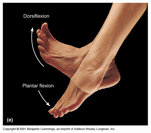
Tibialis anterior lets you bend your foot upwards (dorsiflexion) and also helps to invert the foot (roll outwards) when you walk. The inversion of the foot is an important part of the normal gait pattern.
Flexor hallucis longus is responsible for flexing the big toe, supinating the ankle (turning inwards), and in pointing your foot (plantar flexion).

Restrictions in any of these structures can cause excessive pronation, which in turn leads to hip restrictions, and subsequent knee problems. Obviously, in such situations, treating just the structures of the knee will not resolve the knee problem. Instead, the practitioner must treat the knee, and then, based on the biomechanical and hands on analysis, treat all other affected structures in the knee's kinetic chain. The knee problem will only be resolved when restrictions in all these affected structures are removed and any abnormal muscle firing patterns are addressed with exercise.

Abnormal Muscle Firing Patterns

A key point here is that for every restriction that is created an altered muscle firing pattern will also be created. These dysfunctional movement patterns will still remain after the restriction (adhesion/scar tissue) has been removed. Only a corrective program of exercises will re-establish a normal motion pattern. That is why it is essential to combine both the removal of the adhesion with the exercise protocols, both are key components.

If you would like more information or to purchase our books please go to www.releaseyourbody.com . 

If you would like information about our clinic in Calgary Alberta please go to www.kinetichealth.ca.

(COPYRIGHT KINETIC HEALTH 2012 – ALL RIGHTS RESERVED)